Causes Of Abnormal Bone Marrow On Mri
As with all cancers the earlier its caught the better. Bone marrow serves a crucial function for the body producing bone marrow stem cells and blood products.
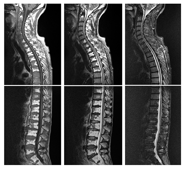
Diffuse Appearance Of Red Bone Marrow On Mri Mimics Cancer Metastasis And Might Be Associated With Heavy Smoking
The presence of BME is an unspecific but sensitive sign of.
Causes of abnormal bone marrow on mri. There are two main types of bone marrow and they each perform specific roles. MRI basics Quick hits T1 T1-weighted images are generally considered to show the best anatomy Although they are not that sensitive to pathology They have the best signal-to-noise per-unit time of scanning On T1-weighted images. Cancer can develop anywhere in the body. Metastasis of malignant neoplasms to bone is common with metastases being far more prevalent than primary bone malignancies12Indeed bone is the third most common organ affected by metastasis surpassed only by the lungs and liver2-4 and is the most common site of distant metastasis from primary breast carcinomaOver the past. Bone lesions are lumps or masses of abnormal tissue produced when cells within the bone start to divide uncontrollably. When cancer is detected in bones it either.
The bone marrow is one of the largest organs in the body and is visible in every magnetic resonance MR imaging study. MRI finds bone and bone marrow disorders. If only an aspiration is to be done it may be taken from the sternum breast bone. The term is a misnomer as the lesion is neither an aneurysm nor a cyst. They can stem from an injury or infection and they may result in bone tumors. Learn more about the symptoms causes diagnosis and treatment of leukemia.
The abnormal cell multiplies rapidly. A tumor may be malignant which means its growing aggressively and spreading to. These blood cells are not fully developed and are called blasts or leukemia cells. Causes vary and treatment is dependent upon the type of fracture. MRI works better than X-rays to pick up signs. Then if you do have that catching or locking sensation that could be potentially a be tear of the meniscus that causes the meniscus to become out of place or flipped such as a flap tear or bucket1handle tear.
This article will provide a systematic overview of the most common disorders in the ankle and foot associated with BME. The process of the bone marrow creating red blood cells white blood cells and platelets is called hematopoiesis. The disease can lead to weakened bones anemia abnormal kidney function and other health problems. Leukemia is a cancer of the blood and bone marrow. Read about types of bone fracture broken bones. These samples can help to determine the percentage of plasma cells and when tested in the lab they can aid in identifying whether the abnormal plasma cells are producing kappa or lambda light chains.
Body magnetic resonance imaging MRI. Bone marrow cancer is a broad category that includes many types and treatment options. It generally presents with pain and swelling in the affected bone. Abnormal synovium or the lining of the knee can be inflamed. Bone marrow edema BME is one of the most common findings on magnetic resonance imaging MRI after an ankle injury but can be present even without a history of trauma. It also can help assess iron concentration in the heart liver and other organs.
Symptoms may include bleeding and bruising bone pain fatigue fever and an increased risk. Bone cancer is a malignant tumor that arises from the cells that make up the bones of the body. In the bone marrow myeloma cells crowd out healthy blood cells leading to fatigue and an inability to fight infections. Aneurysmal bone cyst ABC is a non-cancerous bone tumor composed of multiple varying sizes of spaces in a bone which are filled with blood. Multiple myeloma is a rare condition that causes cancerous plasma cells to be produced multiply and build up in the bone marrow. Bone cancer occurs when a tumor or abnormal mass of tissue forms in a bone.
Plasma cells are a type of white blood cell found in the bone marrow. This abnormal watery material within the bone marrow results from the leakage of fluid and blood into the bone due to damage to the walls of surrounding. Categorization of Bone Marrow Lesions. The damage may not be visible for 2 weeks on an X-ray so the more detailed scans are more effective for recent injuries. It is composed of a combination of hematopoietic red marrow and fatty yellow marrow and its composition changes throughout life in response to normal maturation red to yellow conversion and stress yellow to red reconversion. They are part of the immune system and help fight infection.
The most common broken bones are stress fractures rib fractures skull fractures hip fractures and fractures in children. Myeloma is a type of blood cancer that develops from plasma cells in the bone marrow. These abnormal cells then spill into the bloodstream. Pressure on neighbouring tissues may cause compression effects such as neurological symptoms. This is particularly useful in patients with multiple blood transfusions and concern for iron overload. Based on the type and relative proportion of signal alterations on conventional T1W and T2W MRI images various etiologies of bone marrow lesions can conveniently be divided into three categories Table 1Category I includes bone marrow lesions related to trauma insufficiency injury aseptic necrosis biomechanical disusecomplex.
The samples are usually taken from the back of the pelvic hip bone but sometimes other bones are used instead. Bone marrow edema also referred to as a bone marrow lesion is a condition where the normal fatty bone marrow is replaced with a watery material when there is damage to normal bone structure. In leukemia this rapid out-of-control growth of abnormal cells takes place in the bone marrow of bones. Tissues with short T1 times like subcutaneous fat or fatty bone marrow appear bright Tissues with long T1 times like fluid cotical bone appear. Because cancer cells dont mature and then die as normal cells do they accumulate eventually overwhelming the production of healthy cells. Leukemia is a type of blood cancer that affects your bone marrow which makes blood.
For a bone marrow aspiration you lie on a table either on your side or on your belly. Meniscal tear could be a cause. An X-ray MRI or CT scan can reveal any bone damage. See the Magnetic Resonance Imaging MRI Safety page for more information about. Leukemia also spelled leukaemia and pronounced l uː ˈ k iː m iː ə loo-KEE-mee-ə is a group of blood cancers that usually begin in the bone marrow and result in high numbers of abnormal blood cells. In simple terms cancer is defined as the uncontrolled growth of abnormal cells.
Myeloma is often called multiple myeloma because most people 90 have multiple bone lesions at the time it is diagnosed. Your doctor may also take an MRI scan of your spine and pelvis to look for any lesions or damage. This is also known as primary bone cancerPrimary bone tumors are tumors that arise in the bone tissue itself and they may be benign or malignant bone cancerBenign non-cancerous tumors in the bones are more common than bone cancers. A bone marrow biopsy takes a small amount of bone as well as the marrow cells and fluid from inside the bone. Abnormal synovial fluid within the joint.
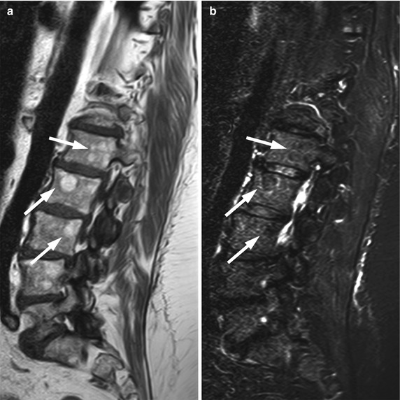
Mri Of The Abnormal Bone Marrow Focal Pattern Radiology Key

Bone Marrow Edema An Overview Sciencedirect Topics
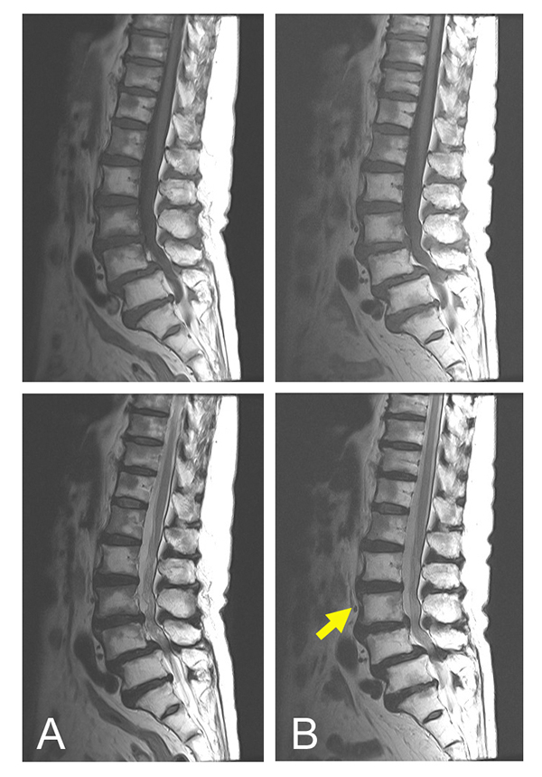
Diffuse Appearance Of Red Bone Marrow On Mri Mimics Cancer Metastasis And Might Be Associated With Heavy Smoking
/GettyImages-1141176554-6bf8f71a554c42518de57c6b999196a4.jpg)
Bone Marrow Edema In The Knee Causes Symptoms Treatment
Mr Imaging Characteristics Of Cranial Bone Marrow In Adult Patients With Underlying Systemic Disorders Compared With Healthy Control Subjects American Journal Of Neuroradiology

Bone Marrow Edema Aids Diagnosis And Prognosis
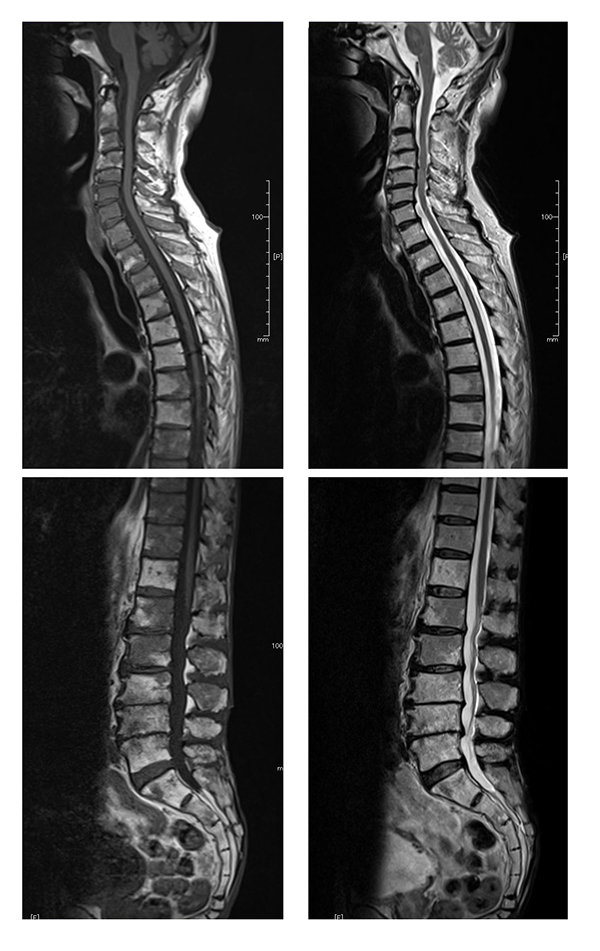
Diffuse Appearance Of Red Bone Marrow On Mri Mimics Cancer Metastasis And Might Be Associated With Heavy Smoking
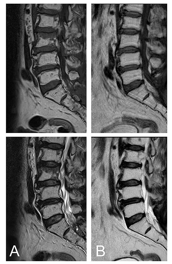
Diffuse Appearance Of Red Bone Marrow On Mri Mimics Cancer Metastasis And Might Be Associated With Heavy Smoking
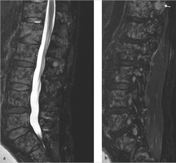
28 Diffusely Abnormal Marrow Signal Within The Vertebrae On Mri Radiology Key

Evaluating The Varied Appearances Of Normal And Abnormal Marrow Radsource
28 Diffusely Abnormal Marrow Signal Within The Vertebrae On Mri Radiology Key

Evaluating The Varied Appearances Of Normal And Abnormal Marrow Radsource
Posting Komentar untuk "Causes Of Abnormal Bone Marrow On Mri"