Si Joint Inflammation Mri
MAGNETIC RESONANCE IMAGING OF SACROILIAC JOINT INFLAMMATION H. In the adult ASAS criteria there is a clear definition of a positive MRI for sacroiliitis 15.

Mri Lesions In The Sacroiliac Joints Of Patients With Spondyloarthritis Youtube
In patients pts with non-radiographic axial SpA nr-axSpA there may be inflammation along the spine in the absence of sacroiliac joint SIJ inflammation on MRI.

Si joint inflammation mri. These two joints are located where the sacrum the triangular last section of the spine meets the ilium a part of the pelvis. Sacroiliitis is the inflammation of one or both of your sacroiliac joints. HALLGREN A consecutive series of 27 patients with symptoms compatible with sacroiliitis underwent magnetic reso- nance imaging MRI of the sacroiliac joints. In summary MRI allows for earlier detection of the joint synovitis erosions and bone marrow edema that are present in inflammatory arthritis facilitating earlier diagnosis and more prompt institution of appropriate therapy. Neoplasms of bone joint or soft tissue Infections of bone joint or soft tissue Inflammation of the Sacroiliac joint Sacroiliac joint dysfunction ankylosing spondylitis degenerative arthritis Sacroiliac joint pain avascular necrosis rheumatoid arthritis osteoarthritis Sacroiliitis psoriasis gout. This analysis evaluated the existence of spinal inflammation on MRI at baseline BL in nr-axSpA pts with and without inflammation in the SIJs on MRI.
Among patients with baseline SPARCC scores 40 had an SI joint score of 2 and 52 had a spine score of 2. Obstetricians often treat sacroiliac joint pain caused by pregnancy. Findings on MRI were analyzed for correlation with multiple clinical characteristics and measures of disease activity including radiographic scoring. Small field-of-view MRI of the sacroiliac joint imaging seems to confer benefit over large large-field-of-view pelvic MRI for detection of osteitis erosions synovitis and bone sclerosis in pediatric patients. A lot of things can cause SI joint dysfunction including overuse and injuries. Positive evidence of inflammation on MRI was defined as a SPARCC score of 2 for either the SI joints or the spine.
Sacroiliitis is a common source of lower back pain or pain in the buttocks or thighs. Find a Doctor Near You Trained in Diagnosis of the Sacroiliac Joint. Forty-nine percent of patients with baseline SI joint scores of. In adults the presence of active sacroiliitis on MRI is a key criterion in the Assessment of Spondyloarthritis International Society ASAS classification 15 16 17. MRI of the SI joints commonly shows non-inflammatory disease in patients clinically suspected of sacroiliitis. What kind of doctor treats SI joint dysfunction.
Ad Get Your Magnetic Resonance Imaging at The Regions Most Preferred Orthopedic Practice. Central reading decreases measurement error but does not translate easily to what is usually done in clinical. Rheumatologists are experts in treating SI joint pain caused by inflammatory arthritis such as ankylosing spondylitis psoriatic arthritis reactive arthritis rheumatoid arthritis and gout as well as SI joint pain from other causes. Ad Is It Your SI Joint. We obtain all sacroiliac joint imaging with a FOV of 160 mm craniocaudal 200 mm wide a slice thickness of 4 mm and an interslice gap of 08 mm. Other MRI features representing active inflammation of the SI joint such as enthesitis or capsulitis alone are not sufficient for a positive MRI for sacroiliitis.
Structural postinflammatory lesions in the sacroiliac joints such as sclerosis fat infiltration erosion or ankyloses are not included in the definition 15 16 17. An MRI is helpful for looking at the tissue surrounding the joint to see if the SI joint inflammation is caused by a mobility or stress issue or if there may be an underlying autoimmune disorder causing the problem. The method has been shown to have both high sensitivity and high specificity in the assessment of inflammatory. Imaging findings play an important role in the diagnosis of sacroiliitis in patients with ankylosing spondylitis and MRI of the sacroiliac joint has been shown to be able to reveal the anatomy and degree of inflammation without the use of ionizing radiation 6. For 15 to 30 of people with long-term lower back pain one of the sacroiliac SI joints which connect the pelvis with the spine is the source. Call or Request an Appointment Online Today and Get Your MRI When its Convenient for You.
The effect of MRI-detected inflammation on the development of radiographic damage at the sacroiliac joints SIJ level in patients pts with axial SpA axSpA has been previously shown when images were scored by trained central readers 1. Many studies are negative despite inflammation occurring while a great number of studies are inconclusive and of little value to the diagnostic process as a whole. MRI and CT scan evaluation of the joint may or may not be capable of visualizing evidence of sacroiliac joint inflammation. Ad The SI joins or sacroiliac joins are found between the iliac bones and other soft tissue. The diag- nostic sensitivity of MRI was similar to that of computed. The Right Diagnosis Will Lead You to the Right Treatment.
The sacroiliac joints were evaluated by two radiologists for enhancement subchondral bone marrow edema erosions and subchondral fatty marrow infiltration. Our study shows that non-inflammatory disease is more common than true sacroiliitis on MRI of the SI joints in patients with inflammatory type back pain.

Magnetic Resonance Imaging Of The Sacro Iliac Joint Sij A Axial T1w Download Scientific Diagram

Imaging The Patient With Sacroiliac Pain Sciencedirect

Atlas Of Mri Findings Of Sacroiliitis In Pediatric Sacroiliac Joints To Accompany The Updated Preliminary Omeract Pediatric Jamris Juvenile Idiopathic Arthritis Mri Score Scoring System Part I Active Lesions Sciencedirect

Overcoming Two Technical Pitfalls In Mri Of Paediatric And Adolescent Sacroiliitis Clinical Radiology

Pin On Sacro Iliac Workouts Stretches

Atlas Of Mri Findings Of Sacroiliitis In Pediatric Sacroiliac Joints To Accompany The Updated Preliminary Omeract Pediatric Jamris Juvenile Idiopathic Arthritis Mri Score Scoring System Part Ii Structural Damage Lesions Sciencedirect

Atlas Of Mri Findings Of Sacroiliitis In Pediatric Sacroiliac Joints To Accompany The Updated Preliminary Omeract Pediatric Jamris Juvenile Idiopathic Arthritis Mri Score Scoring System Part Ii Structural Damage Lesions Sciencedirect

Common Incidental Findings On Sacroiliac Joint Mri In Children Clinically Suspected Of Juvenile Spondyloarthritis European Journal Of Radiology Open

Common Incidental Findings On Sacroiliac Joint Mri In Children Clinically Suspected Of Juvenile Spondyloarthritis European Journal Of Radiology Open

Sacroiliac Joint Sij Mri In The Coronal Plane In Two Different Download Scientific Diagram

Common Incidental Findings On Sacroiliac Joint Mri In Children Clinically Suspected Of Juvenile Spondyloarthritis European Journal Of Radiology Open
Pin On Tailbone Pain Doctor
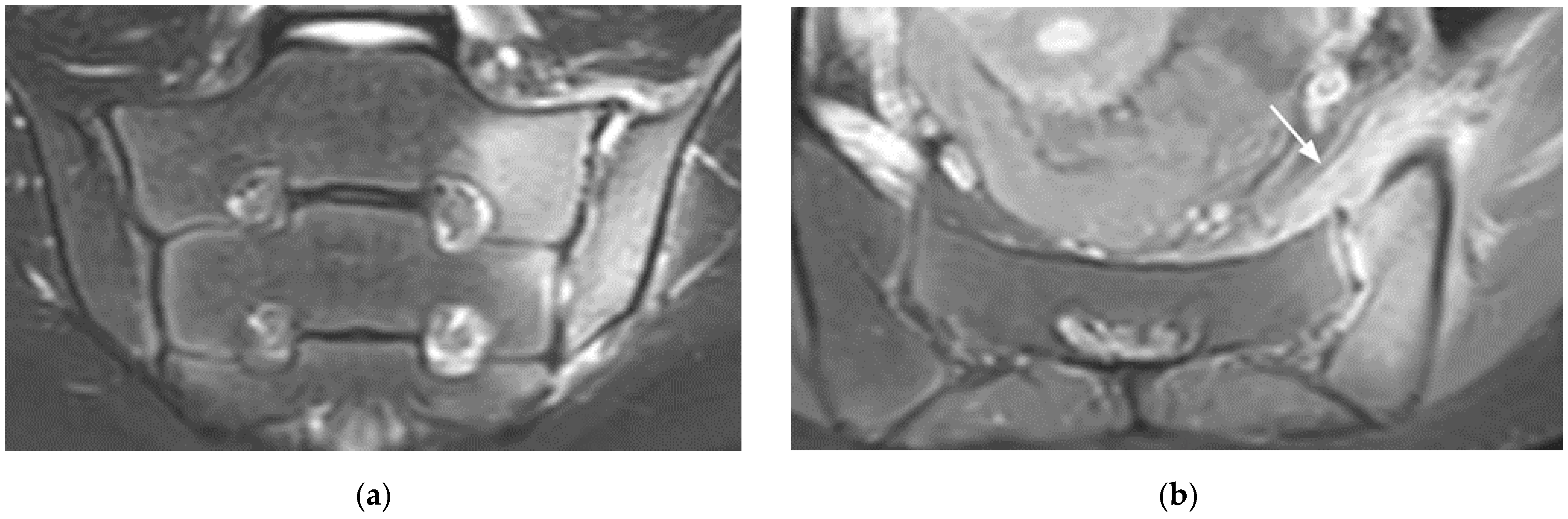
Diagnostics Free Full Text Main Diagnostic Pitfalls In Reading The Sacroiliac Joints On Mri Html
2
Posting Komentar untuk "Si Joint Inflammation Mri"