Spinal Stenosis Mri With Or Without Contrast
A spinal cord injury SCI is damage to the spinal cord that causes temporary or permanent changes in its function. Quantitative parameters of magnetic resonance imaging cannot predict human epidermal growth factor receptor 2 HER2 status in rectal cancer.

Healthcare Extreme How To Read An Mri Lumbar Spine In 8 Easy Steps
Symptoms may include loss of muscle function sensation or autonomic function in the parts of the body served by the spinal cord below the level of the injury.
Spinal stenosis mri with or without contrast. Evaluation of a patient with signs or symptoms of spinal stenosis where MRI or CT are equally appropriate. Because PILD is performed without general anesthesia it may be an option for some people with high surgical risks from other medical problems. Injects contrast dye into the spinal fluid space cerebrospinal fluid to outline the nerves and spinal cord and show evidence of any pressure. The procedure removes a portion of the vertebra to ease pressure on the spinal. MRI basics Quick hits T1 T1-weighted images are generally considered to show the best anatomy Although they are not that sensitive to pathology They have the best signal-to-noise per-unit time of scanning On T1-weighted images. This may be caused by arthritic overgrowth of the facet joints degeneration of the disc with loss of tension in the disc and loss of disc height overriding of the facet joints with concurrent bulging of.
Cervical spine MRI without contrast should be performed. It may be performed with a dye or without a dye. A low correlation exists between the degree of morphological stenosis observed in MRI and clinical symptoms. A CT scan provides excellent definition of the bones while an MRI allows your physician to see the soft tissue structures such as muscles spinal nerves and spinal cord. Term for various functional disturbances or pathological changes in the spinal cord. This International journal Journal of Clinical Neuroscience publishes articles on clinical neurosurgery and neurology and the related neurosciences such as neuro-pathology neuro-radiology neuro-ophthalmology and neuro-physiology.
Injury can occur at any level of the spinal cord and can be complete with a total loss of sensation and. The journal has a broad International perspective and emphasises the advances occurring in Asia the Pacific Rim region Europe. Term for the narrowing of the vertebral canal nerve root canals or intervertebral foramina of the lumbar spine that is caused by encroachment of bone. Magnetic resonance imaging MRI. MRI is safe and painless. Combining DBT with CEDM has been shown to limit the effect of surrounding soft tissue and achieving higher contrast between malignancy and surrounding tissue 2.
The advantage of MRI over X-Ray is that intervertebral discs and nerves are clearly visible in an MRI scan. MRI Abdomen with and without contrast MRI Pelvis with and without contrast 74183 72197 Crohns disease Crohns fistulous disease Celiac disease Sprue Small bowel tumor Yes and glucagon Body BREAST MRI MRI Breast Bilateral with. When you undergo a contrast MRI a contrast injection such as gadolinium or iodine is given. An MRI is a very useful tool for helping your doctors see images of the inside of your body including tissue that cant be seen on a conventional x-ray. Gadolinium MRI contrast injections improve diagnostic accuracy in some conditions such as inflammatory and infectious diseases of the brain spine soft tissues and bones by making images clearer so that the radiologist can. MRI can also detect soft tissue anomalies like spinal cord tumours abscesses or bony spurs.
MR Lumbar without contrast Disc bulges Disc herniation Incontinence Low back pain Radiculopathy Spinal stenosis Trauma. A dye is a contrast agent that is given to you by the IV route. Tissues with short T1 times like subcutaneous fat or fatty bone marrow appear bright Tissues with long T1 times like fluid cotical bone appear. Relieve lumbar spinal stenosis. Types of scans. Contrast material is often not required to evaluate for cervical foraminal stenosis.
MRI with and without contrast. Computed tomography Magnetic resonance imaging Ultrasonography Digital radiology Interventional radiology. Cervical neural foraminal stenosis was classified into four grades according to MRI findings on T 2 weighted oblique sagittal images Figure 1The grading system used was modified from a pre-existing grading system for lumbar neural foraminal stenosis suggested by Wildermuth et al Grade 0 refers to the absence of. If the narrowing is substantial it can cause compression of the spinal cord or spinal nerves. To diagnose spinal stenosis your doctor may ask you about signs and symptoms discuss your medical history and conduct a physical examination. Creates images by using powerful magnets and computer technology and can show the spinal cord nerve roots and surrounding areas as well as enlargement degeneration and tumors.
But metal in the scanner can cause serious safety problems or reduce the quality of the images. Magnetic Resonance Imaging MRI. MRI is useful for diagnosing lumbar spinal stenosis and identifying the degree of degenerative change and the size of the spinal canal 1 24 25. Digital mammography spawned the development of several derived technologies including digital breast tomosynthesis DBT and contrast-enhanced digital mammography CEDM 1. MRI is generally not indicated if. A contrast agent is a liquid injected into your body to make certain tissues clearly visible during the imaging process.
Lumbar spinal stenosis is a condition where the spinal canal central stenosis or one or more of the lumbar vertebral foramina foraminallateral stenosis becomes narrowed. With and without contrast which is the best imaging for detecting spinal infections. MR Lumbar without contrast with Flexion Extension Back painlower extremity radicular symptoms especially when position dependent. However MRI is currently by far the most commonly used imaging method for the ac-curate evaluation of spinal canal stenosis. There are two types of MRI imagingMRIs with and without contrast. MRI visualizes not only the width and length of the spinal canal but also depicts in detail the spinal cord intervertebral disks osteo-phytes and ligaments all of which are po-tential causes of spinal canal stenosis 2.
MRI grading system for cervical neural foraminal stenosis. Before your exam its very important to fill out the safety screening form carefully. MRICT showing at least moderate central canal spinal stenosis neuro-radiologist definition with symptoms correlating with stenosis symptoms of backbuttockleg pain without unilateral radicular pain in a neurogenic claudicatory fashion relieved by stooping or sitting down. Incidence of pulmonary embolism and impact on mortality in patients with malignant melanoma. Clinical Radiology is published by Elsevier on behalf of The Royal College of RadiologistsClinical Radiology is an International Journal bringing you original research editorials and review articles on all aspects of diagnostic imaging including. For evaluation of recurrent symptoms after spinal surgery MRI with and without gadolinium enhancement is the preferred method of imaging.
Often refers to nonspecific lesions in contrast to the inflammatory lesions. In contrast to the back and leg pain associated with degenerative lumbar stenosis the pain associated with a lumbosacral spinal tumor typically worsens with recumbency awakens the. Lateral Recess Stenosis is a condition where the narrowing reduces the available space within the exit doorway foramen of the spinal canal.

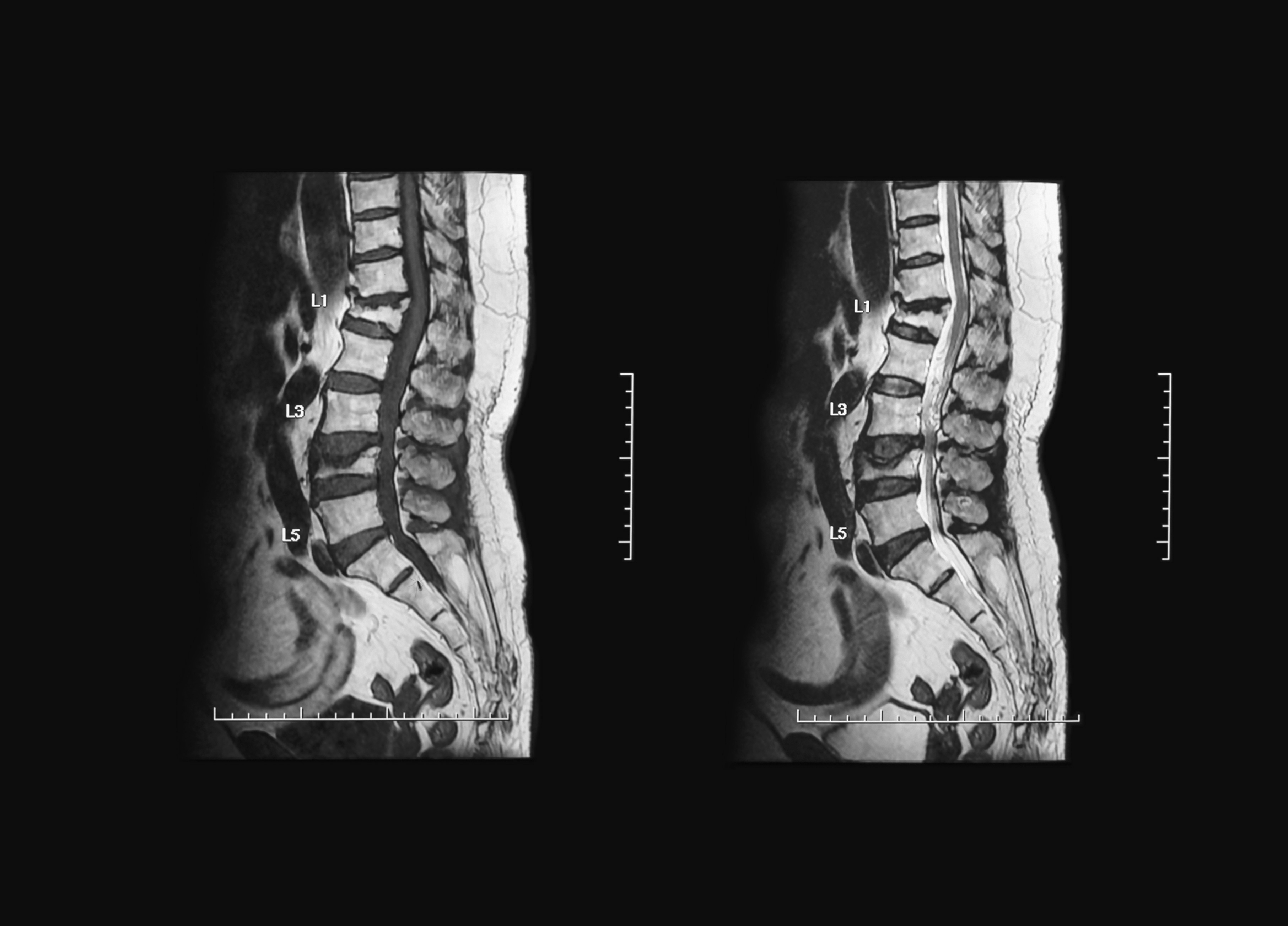
Preoperative Mri Of The Lumbar Spine Without Intravenous Contrast And Download Scientific Diagram
Ge Healthcare United States
Racgp Making Sense Of Mri Of The Lumbar Spine
Delayed Post Traumatic Spinal Cord Infarction In An Adult After Minor Head And Neck Trauma A Case Report Journal Of Medical Case Reports Full Text
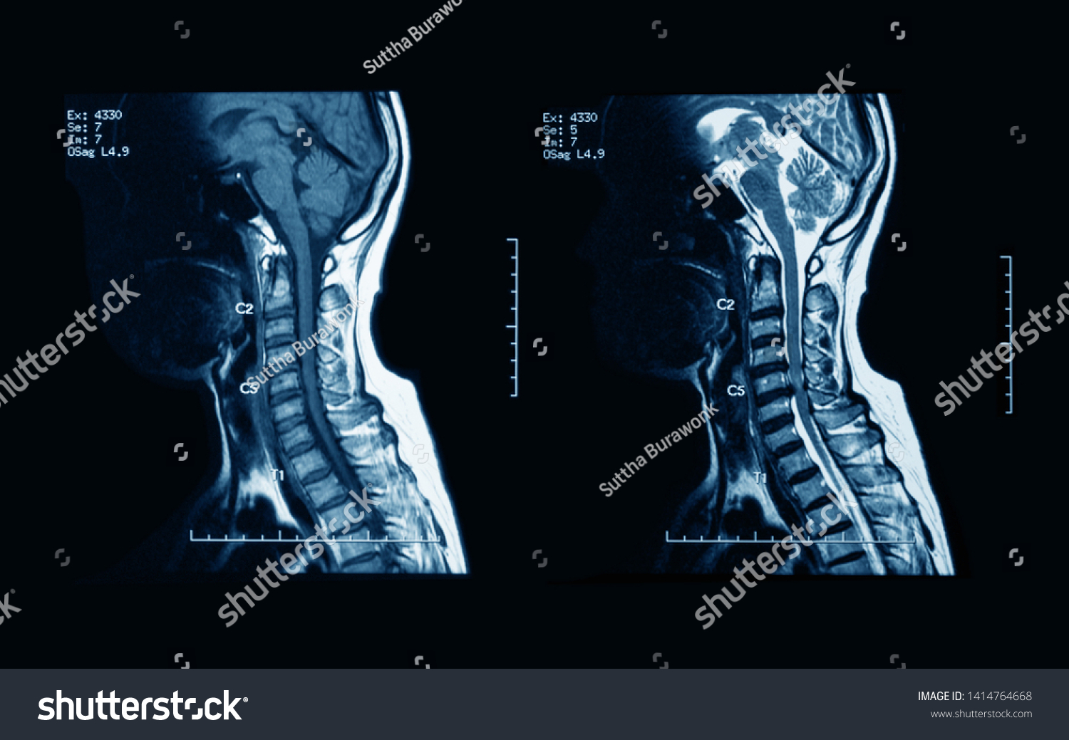
Mri Scans Cervical Spine Without Contrast Stock Photo Edit Now 1414764668

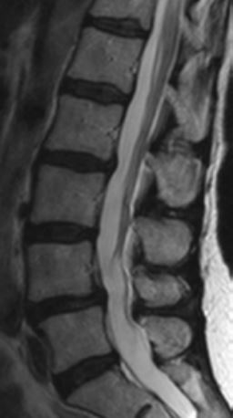
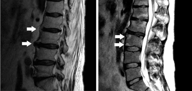
Lumbar Magnetic Resonance Imaging Without Contrast Showing Mild Download Scientific Diagram
Spine Mri Quick Reference Guide For Physicians

T1 Weighted Magnetic Resonance Imaging Mri Without Contrast Of Whole Download Scientific Diagram

Imaging Of The Spine And Spinal Cord An Overview Of Magnetic Resonance Imaging Mri Techniques Sciencedirect

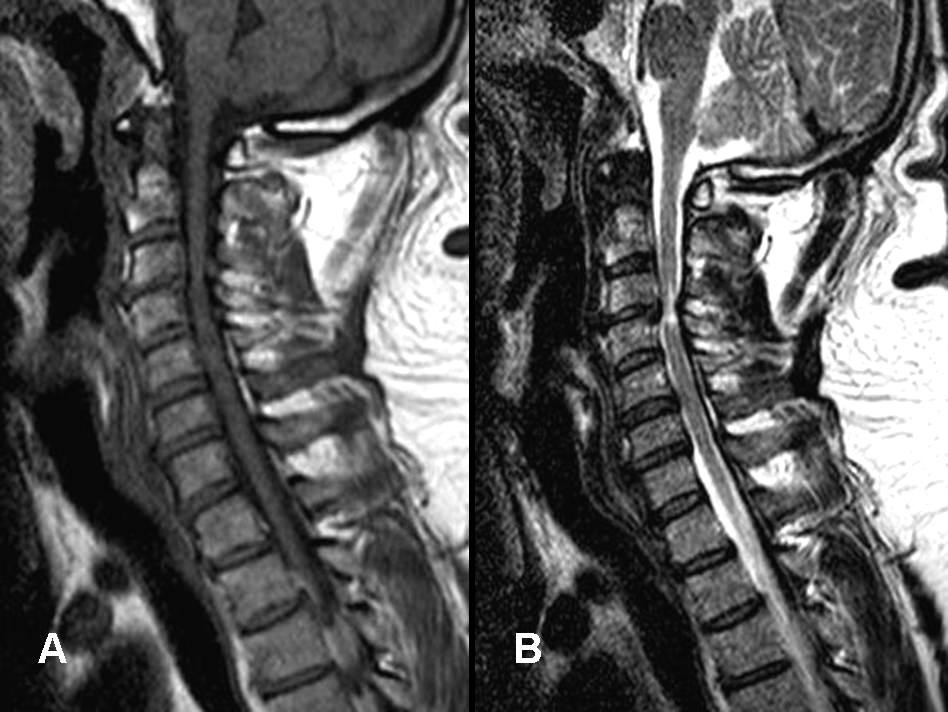
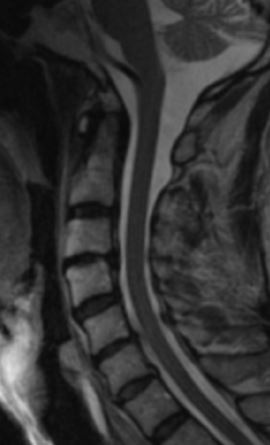
Magnetic Resonance Imaging Mri Without Contrast Of The Cervical Spine Download Scientific Diagram

Mri Cervical Spine Without Contrast Showing Mild Multisegmental Download Scientific Diagram
Spine Mri Quick Reference Guide For Physicians
2

Noncontrast Mri Cervical Spine Search Pattern Youtube
Posting Komentar untuk "Spinal Stenosis Mri With Or Without Contrast"