Mri Of Thigh What Does It Show
Indications for mri thigh scan Marrow abnormalities. A leg MRI also creates pictures of the knee.

Leg Mri Two Views
Magnetic Resonance Imaging MRI of Thoracic Spine.

Mri of thigh what does it show. The dye is considered to be safe but may be harmful to those undergoing dialysis. MRI does not use radiation x. What is the CPT code for MRI of the thoracic spine. Radiologists need to be familiar with typical MRI findings in order to accurately detect and classify muscle injuries. This may include the ankle foot and surrounding tissues. MRI of Upper Leg Femur Magnetic Resonance Imaging MRI is a safe as well as painless procedure.
MRI is the imaging test of choice for evaluating muscle and tendon disorders. Radiography is a relatively inexpensive means of screening patients for heterotopic ossification avulsion fractures and other osseous injuries. Depending on what your doctor is looking for this test may be ordered with or without IV contrast. Obviously if there are degenerative elements in the hip loss of cartilage bone on bone destruction hip instability causing soft tissue structural damage there will be pain. In my practice it is not uncommon for an MRI study to be performed for something palpable but no. An MRI or Magnetic Resonance Imaging is a scan that is able to render images of soft tissue structures throughout the body.
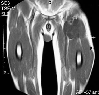
BR37ZZZ is a valid billable ICD-10 procedure code. Procedure for MRI Scan - Thigh Screening Test. In case of Acute trauma MRI is done to check for the possible injuries Ligament tear. Discover all of the pertinent medical information regarding magnetic resonance imaging. Obviously it is critical to evaluate the MRI images personally when making a diagnosis so I cant give you an answer specific to your case. Another lesion of increased signal intensity is seen in the gluteus maximus muscle arrowhead.
A leg MRI magnetic resonance imaging scan of the leg uses strong magnets to create pictures of the leg. Hip pain may require a test called magnetic resonance imaging to diagnose the underlying cause of the discomfort. MRA uses magnetic resonance imaging MRI to take images of blood vessels. B Corresponding axial T2weighted MR image shows a mature ossifying lesion of low signal intensity adjacent to the proximal femur arrow and a high signal intensity mass in the surrounding soft tissue arrowhead. This allows physicians to view a patients full spinal anatomy in order to determine the cause of a patients pain which can then be correlated to symptoms to provide a diagnosis. MRIs for Diagnosing Nerve Pain.
Myositis MRI muscles Rheumatology key messages. MRI uses a magnetic field and radio waves to create detailed images of the organs and tissues within your body without the use of ionizing radiation. During a hip MRI the patient is asked to lie very still in a tube-like structure as the test is performed. The doctor orders an MRI the MRI reveals nothing. Ad Learn the uses and side effects of magnetic resonance imaging MRI scans today. A leg MRI scan may be done to check for certain cancers or other illness.
A MRI scan of the leg can identify the cause for decreased motion or increased pain of a particular joint. Muscle injuries of the hip and thigh are a highly relevant issue in competitive sports imaging. Contrast MRI is also used to detect Mortons neuroma in the leg. MRI accurately documents the extent and intensity of the muscle abnormalities. One of the best reasons to get a magnetic resonance scan is that the surgeon may use it as a kind of map for surgery. The gold standard in diagnostic imaging of muscle injuries is magnetic resonance imaging MRI.
Magnetic resonance angiography MRA of the legs showing blood vessels. Answer 1 of 3. The diagnosis is based on a typical clinical presentation elevated serum skeletal muscle enzymes and findings on electromyography and muscle biopsy. The dye temporarily binds to certain tissues and then is gradually eliminated through urine. To detect the Ulcers. CT provides for cross-sectional.
Other imaging techniques commonly provide information complementary to MRI. The MRI may show tissue that has cancer cells and tissue that does not have cancer cells. MRI of the thigh muscles shows a distinct pattern of oedema and fatty infiltration and can be used to monitor the treatment of patients with anti-SRP myopathy. For example an MRI may show that a tumor is wrapped around an important nerve or blood vessel which would. Proper interpretation of the findings is crucial especially in. A leg MRI also creates pictures of the knee.
Strains partial and complete tears tendonitis tendonopathy. A loud clanging noise may be heard throughout the test as the magnets move to provide accurate imaging. To scan any Fractures and have a more clear and exact view of the injuries caused. Most healthcare practitioners use gadolinium as the contrast dye for MRI. A MRI scan of the leg can show healthcare providers how well a treatment for a disease is working and the. It is also done to check the Infections of bone joint or soft tissues.
A Femur MRI magnetic resonance imaging scan also known as Thigh or Femur uses strong magnets to create pictures of the leg. Avascular necrosis marrow edema syndromes and stress fractures Muscle and tendon disorders. Polymyositis is a rare autoimmune and sometimes paraneoplastic inflammatory myositis.

An Mri Scan Of His Pelvis Left And Right Thigh With And Without Download Scientific Diagram

Mri Thigh Upper Legs Planning Mri Hamstring Protocols Indications For Mri Thigh Scan

Mri Thigh Upper Legs Planning Mri Hamstring Protocols Indications For Mri Thigh Scan
Mri Of Thigh Abscess In The Vastus Medialis Muscle Is Shown Download Scientific Diagram
Which Mri Findings Are Characteristic Of Liposarcoma

T1 Weighted Mri Images Of Thighs And Legs From 3 Fshd1 Patients Download Scientific Diagram

Muscle Mri For Neuromuscular Disorders Practical Neurology
%20positioning.jpg)
Mri Thigh Upper Legs Planning Mri Hamstring Protocols Indications For Mri Thigh Scan

Mri Thigh Upper Legs Planning Mri Hamstring Protocols Indications For Mri Thigh Scan

Magnetic Resonance Imaging Mri Cure Ibm

Supplemental Materials For Normal Mr Imaging Anatomy Of The Thigh And Leg Magnetic Resonance Imaging Clinics

Example Of Mri Slice Mid Thigh With The Knee Extensors And Flexors Download Scientific Diagram

Figure 4 From Normal Mr Imaging Anatomy Of The Thigh And Leg Semantic Scholar

Mri Of Right Thigh A Large Mass Lesion Measuring 18 8 7 5 Cm In The Download Scientific Diagram
Posting Komentar untuk "Mri Of Thigh What Does It Show"