Pituitary Macroadenoma Mri Findings
Elevated prolactin may occur in several situations. Benign adenoma invasive adenoma and carcinomasMost adenomas are benign approximately 35 are invasive and just 01 to 02 are carcinomas.
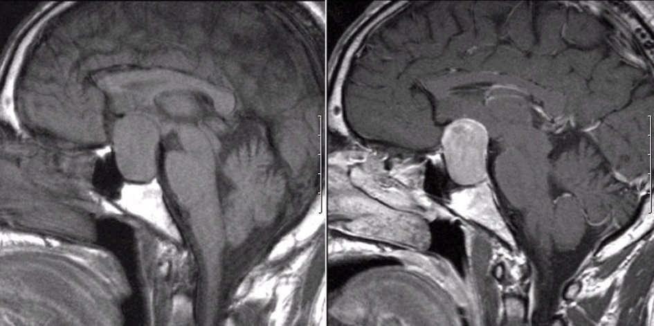
Pituitary Macroadenoma And Periventricular Leukomalacia Radiology Case Radiopaedia Org
72 patients were excluded and 146 participants were enrolled into the run-in phase.
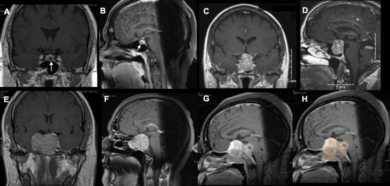
Pituitary macroadenoma mri findings. A pituitary tumor is a growth of abnormal cells in the tissues of the pituitary gland. Pituitary adenomas PA are non-metastasizing neoplasms that arise in the pituitary gland. Non-invasive Detection of Breast Cancer in Lymph Nodes. 12 Typical imaging findings of an uncomplicated pituitary adenoma include slow enhancement compared with that of the pituitary gland lateral deviation of the infundibulum and isointense. A 70-year-old female with pituitary gland macroadenoma. Pituitary lesions albeit relatively infrequent can significantly alter the quality of life.
For example in a. 50 91 of 55 participants who. Even if an X-ray MRI and Bone scan are of the same patient each is marked separately. Since January 2014 all hospitalizations and outpatient treatments eligible for Philhealth coverage are already being covered under the Philhealth Case Rate system. A pituitary adenoma may be suspected based on symptoms the medical history and physical findings of acromegaly. Pituitary adenomas represent from 10 to 25 of all.
The onset of new or worsening headache or a change in vision or both mandates the urgent performance of formal visual field testing and a pituitary MRI without the use of gadolinium. D Visual field left eye 042018 at initial visit. E OCT of optic disc with RNFL. Once your doctor suspects Cushings disease based on clinical findings and hormonal testing a magnetic resonance imaging MRI scan of the pituitary gland is the best way to detect the presence of an adenoma in Cushings disease. B Optic disc left eye. Previously PAs were classified based on their sizes.
Fluorescent and MRI Targeted Probes for the Melanocortin Receptor 1 on Melanomas and Micelle Complexes for Drug Delivery 10MA024 Molecular Imaging Probe. 92 participants were randomly assigned to oral octreotide n55 or iSRL n37. A blood prolactin level is necessary to determine if an elevated prolactin level is a prolactinoma in relation to the pituitary MRI findings. Damage to the gland can impair any or all of the six hormones secreted by the pituitary gland and can lead to complex issues that can cause health issues in many areas of the bodyPrior to surgery on the pituitary gland you can expect to have a CT scan MRI or possibly both done to evaluate the size and shape of the gland and the tumor. Those smaller than 10 mm were known as microadenoma and the rest as macroadenoma. A Optic disc right eye.
Damage to the gland can impair any or all of the six hormones secreted by the pituitary gland and can lead to complex issues that can cause health issues in many areas of the bodyPrior to surgery on the pituitary gland you can expect to have a CT scan MRI or possibly both done to evaluate the size and shape of the gland and the tumor. For medical cases 30 of the fixed amount is for Doctors Professional Fees PF and 70 is for hospital costs. Explaining your symptoms is a crucial part of diagnosis as your doctor uses the information to determine whether a pituitary tumor is secreting an excess of hormones and if there is evidence of pituitary insufficiency. This article highlights the role of advanced imaging modalities in evaluating pituitary-hypothalamic axis lesions. 10MA069N Molecular Imaging Probe. Each medical or surgical case is covered by a fixed amount.
Pituitary adenoma is a benign neoplasm that arises from the adenohypophysis and is the most common intrasellar pathology accounting for 1015 of all intracranial neoplasms. Between Feb 11 2016 and Aug 20 2020 218 patients were assessed for eligibility. Updated June 2 2014. Medications that cause a high prolactin usually a normal MRI of the pituitary a growth in the pituitary that causes small elevation of. So make sure you state all your findings for each. Practice as much as possible with a friend or faculty and by presenting aloud.
Pituitary tumors form in the pituitary gland a pea-sized organ in the center of the brain just above the back of the noseThe pituitary gland is sometimes called the master endocrine gland because it makes hormones that affect the way many parts of the body work. However in patients with macroadenoma who did not undergo surgery or irradiation before pregnancy the risk of symptomatic pituitary tumor enlargement was 31. In 1773 John Fothergill was the first to fully describe trigeminal neuralgia in an article presented to the Medical Society of London titled On a Painful Affliction of the FaceIn 1829 Charles Bell distinguished the specific functions of the trigeminal and facial nerves and introduced the idea that the paroxysmal pain in trigeminal neuralgia is directly related to nerve. In recent years the proteomic studies of PAs have led to a better appreciation of the different hormonal status shown. In addition liver metastasis or invasion into the vena cava may be demonstrated in dogs with adrenal carcinomas. 116 patients completed the run-in phase and 30 participants discontinued treatment.
Sella imaging with MRI or CT is done to exclude a pituitary macroadenoma or other mass in men with any of the following. Pituitary adenomas are tumors that occur in the pituitary glandPituitary adenomas are generally divided into three categories dependent upon their biological functioning. MRI detects a pituitary adenoma in about 70 percent of cases. C Visual field right eye 042018 at initial visit. Age 60 years with no other identified cause for hypogonadism Very low total testosterone levels 200 ngdL 694 nmolL. Magnetic resonance imaging MRI is the examination of choice for evaluating hypothalamic-pituitary-related endocrine diseases.
Lactation requires the presence of estrogen progesterone and most. Take every case as a new one however good or bad your last case has been. Galactorrhea or inappropriate lactation is a relatively common problem that occurs in approximately 20 to 25 percent of women. CT or MRI of the brain or abdominal cavity in dogs that do not suppress on the HDDS may demonstrate unilateral adrenal enlargement 50 pituitary macroadenoma 25 or pituitary microadenoma 25. Prepare a script for common cases.
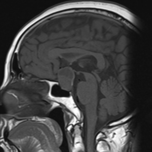
Pituitary Macroadenoma Radiology Case Radiopaedia Org
Pituitary Macroadenoma Mri Online
Pituitary Macroadenoma Mri Online
Surgical Treatment Of Pituitary Adenomas Endotext Ncbi Bookshelf
Pituitary Adenoma3

Macroadenoma An Overview Sciencedirect Topics
Pituitary Macroadenoma Radiology Cases

Pre Operatory Mri Showed A Pituitary Macroadenoma 5 3 X 4 0 X 3 5 Cm Download Scientific Diagram

Mri Pituitary Macroadenoma Stock Image C043 3144 Science Photo Library
Pituitary Macroadenoma Clinical Mri

Pituitarymacro4
Pituitary Adenoma4
Pituitary Macroadenoma With Cystic Degeneration Radiology Case Radiopaedia Org

Mri Of Pituitary Macroadenoma Coronal T1weighted Post Gadolinium Download Scientific Diagram
Posting Komentar untuk "Pituitary Macroadenoma Mri Findings"