Mri Before And After Spinal Decompression
Because an MRI displays detailed images of the organs. The aim of this study was to demonstrate morphological changes in the cervical nerve roots before and after spinal cord decompression surgery in association with the.
Pin On Hernie

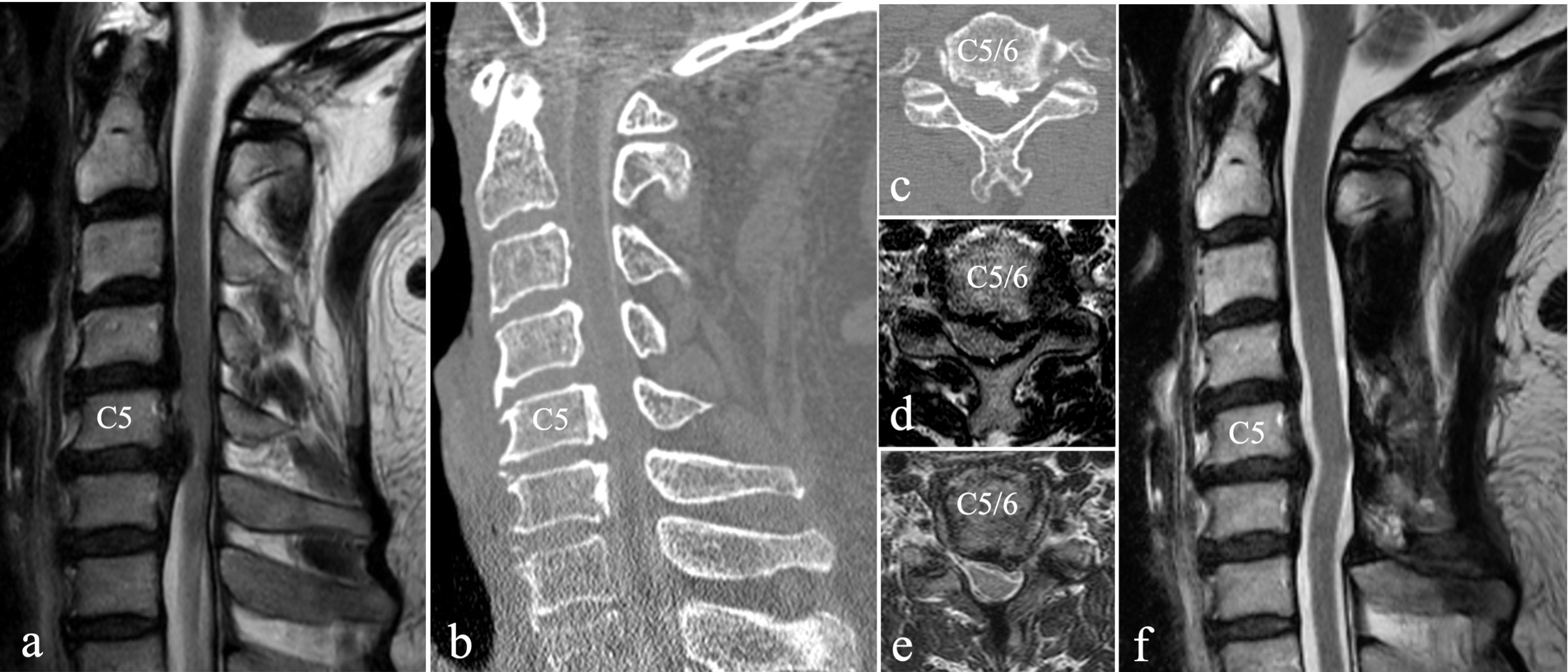
Results and MRI findings before and after decompression surgery.

Mri before and after spinal decompression. Magnetic Resonance Imaging Findings after Treatment with a Non-surgical Spinal Decompression System US MUSCULOSKELETAL REVIEW 2007 51 in intra-discal signal on T2. Patients need to focus on how well they feel after each visit. Discuss risks and benefits of SCS with your doctor. Cervical myelopathy in elderly patients. Decompression surgery for spinal stenosis is elective except in the rare. A resting-state functional MRI.
Be Seen at Mayo Clinic. On the left note that the disc is herniated and compressing the white vertical tissues which are the nerves and spinal cord. Amazingly after completing the treatment the patient reported a pain level of 1. The MRI scan on the left shows a large disc herniation BEFORE Non-Surgical Spinal Decompression treatment. Below are actual before and after MRI images from three patients who chose IDD Therapy to treat their neck back and disc issues. Symptoms should go away after the spinal decompression therapy but to fully relieve the pain between 15 and 30.
Get back to the activities you love. Ad Learn the uses and side effects of magnetic resonance imaging MRI scans today. What can I expect after my MRI scan. Once it has been determined that you are a candidate and we can help you we do everything we can to make sure you are comfortable during each decompression visit so we get the. Alterations of functional connectivity between thalamus and cortex before and after decompression in cervical spondylotic myelopathy patients. Seventy-seven patients underwent anterior decompression and fusion at not more.
Ad Watch a video to learn how spinal cord stimulation can help treat chronic back pain. Spinal decompression therapy was performed by using a lumbar decompression device MAX-D Medirex Korea and the subjects lay on a traction table in the supine position. 1996 Nagata et al. The patient had 22 spinal decompression treatments over a seven-week period. Postoperative changes in the spinal cord in cervical myelopathy demonstrated by magnetic resonance imaging. Diagnostic tests MRI CT myelogram that show stenosis in the central canal or lateral recess.
Nagata K1 Ohashi T Abe J Morita M. This type of spine surgery does carry a reasonable amount of unwanted. The Latest Research Technology Goes into Custom Treatment Plans Tailored Just for You. Ad Better Outcomes from Multidisciplinary Surgical Teams. An average difference of anteroposterior cervical spinal canal distance before and after surgery of CSM was 267 millimetres representing a 40 increase. No simple traction device has ever shown before-and-after MRIs where there was increased disc height or a visible reduction of a DISC HERNIATION.
To the right is an MRI scan of the same patient AFTER Non. Clinical results and MRI findings before and after decompression surgery. On the right after spinal decompression the. Allergic reaction from gadolinium dye is extremely rare. When patients have primarily lower back pain generally the only surgical treatment available is a lumbar spinal fusion. Discover all of the pertinent medical information regarding magnetic resonance imaging.
If a dye injection is used the IV is removed from the arm before you go home. We examined 173 patients with cervical myelopathy of various casuses.
Cureus The Utility Of Augmented Reality In Spinal Decompression Surgery Using Ct Mri Fusion Image

Pin On Back Pain Alternative Treatment Options

Mri Spine Anatomy Free Mri Lumbar Spine Sagittal Cross Sectional Anatomy Anatomy Images Mri Spines

Cureus The Utility Of Augmented Reality In Spinal Decompression Surgery Using Ct Mri Fusion Image
Spinal Stenosis And Chiropractic Care Kempsville Chiropractic Health Articles In 2021 Cervical Spinal Stenosis Stenosis Spinal Stenosis

Pin By Twin Creeks Health On Spinal Decompression Mri Scan Radiology Spinal Decompression

Spinal Decompression In O Fallon Spinal Aid Centers

Preoperative Mri Showing Moderate Spinal Stenosis At The L3 4 Level A Download Scientific Diagram
Cauda Equina Syndrome From Lumbar Disc Herniation Cauda Equina Cauda Equina Syndrome Lumbar Disc

Gonstead Chiropractic Success With Disc Herniation Case Pre And Post Mri Studies Chiropractic Chiropractic Care Chiropractic Marketing

Annotated Color Mri Lumbar Spine Disc Herniation Before After Disk Herniation Mri Lumbar

Lumbar Spinal Mri Of Patient 2 Demonstrating The L5 S1 Discal Cyst Download Scientific Diagram

Post Primary Operative Mri Of 70 Year Old Female With Spinal Stenosis Download Scientific Diagram

Pin On Spinal Injuries


Posting Komentar untuk "Mri Before And After Spinal Decompression"