Carotid Artery Test With Dye
An angiogram is a diagnostic procedure that provides detailed x-ray pictures of your heart and its blood vessels. The reason we call it that is that although it looks normal by angiogram and there is clearly no significant heart blockage there may be deposition of plaque in the walls of the artery that cant be seen on this test.

Pin On Anatomy Physiology
Contrast dye is injected through an IV to illuminate blood vessels in the neck.

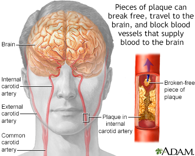
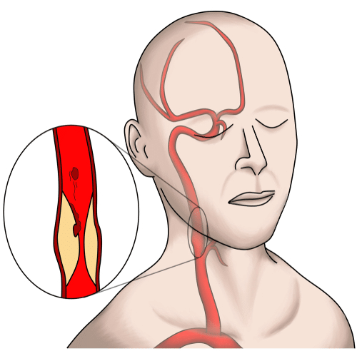
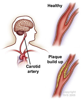
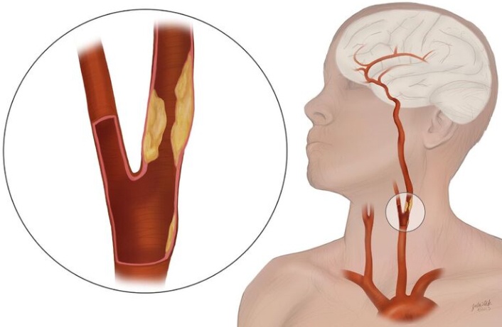
Carotid artery test with dye. Blood vessel walls normally have three layers and a tear in any of these can allow blood to flow into the resulting space causing the vessel to bulge. The artery is repaired with either stitches or a graft. Carotid artery disease is a disease in which a waxy substance called plaque builds up inside the carotid arteries of the neck. Carotid angiography is an invasive imaging procedure that involves inserting a catheter into a blood vessel in the arm or leg and guiding it to the carotid arteries with the aid of a special x-ray machine. This is called digital subtraction angiography DSA. Your doctor may also use a test to diagnose carotid artery disease.
Contrast dye is injected through the catheter while X-rays of the. A carotid artery dissection is a tear in a layer of the wall of a blood vessel called a carotid artery one of two such arteries found in the neck. This is the type of surgery used to open a partly blocked artery. This test is done to assess the blood flow of the carotid arteries. Carotid endarterectomy CEA is surgery to treat carotid artery disease. The scan shows the amount of hardening of the artery wall the disease that causes this hardening is called atherosclerosis.
And it is best determined by having a 4-vessel arteriogram where they place a small catheter in each vesell and. Meta-Analysis Evaluating High-Sensitivity Cardiac Troponin T Kinetics after Coronary Artery Bypass Grafting in Relation to the Current Definitions of Myocardial Infarction. Therefore is not coronary artery disease. A dye is then injected to view the flow of blood from the carotid artery to the brain on a live X-ray monitor. During a carotid endarterectomy your healthcare provider. If contrast is used you may also be asked not to eat or drink anything for 4 to 6 hours before the test.
During this invasive imaging procedure a catheter thin flexible tube is inserted into a blood vessel in the arm or leg and guided to the carotid arteries with the aid of a special X-ray machine. Catheter angiography is considered the gold standard in vascular imaging. An angiogram will show. Sometimes a computer removes the bones and tissues on the images being viewed so that only the blood vessels filled with the dye are seen. This noninvasive painless screening test uses. Angiogram is a minimally invasive test that uses X-rays and a contrast agent injected into the arteries through a catheter in the groin.
That blood flow to your heart is not being restricted by blockages-- a finding that lets you and your doctor know your symptoms are not related to your heart. Carotid endarterectomy is an open surgery to remove the plaque. The material consists of mostly macrophage cells or debris containing lipids calcium and a variable amount of fibrous connective tissueThe accumulated material forms a swelling in the artery wall which may intrude into the lumen of. The coronary artery calcium heart scan is a CT scan of the heart that looks for areas of calcium in the arteries that supply the heart with blood. When the transducer like a microphone is placed on the carotid arteries at certain locations and angles the ultrasonic sound waves move through the skin and other body tissues to the. What is a CT Coronary Artery Calcium Heart Scan.
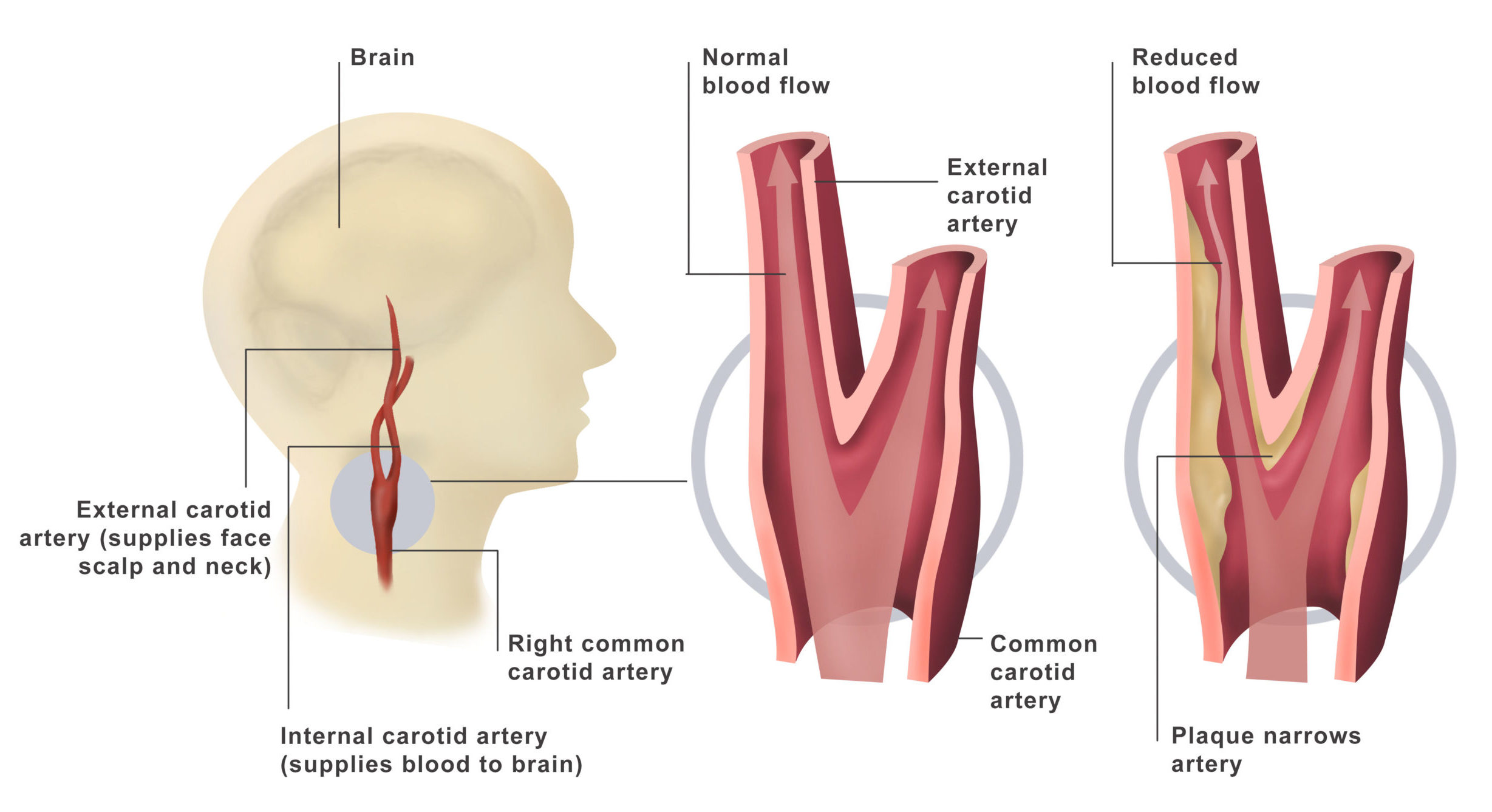
Contrast dye is injected through the catheter so that x-ray movies of your carotid arteries the arteries that supply your brain with. An atheroma or atheromatous plaque plaque is an abnormal and reversible accumulation of material in the inner layer of an artery wall. The carotid artery is in the neck and provides blood to the brain not the heart. Contrast helps certain areas show up better on x-rays. A probe called a transducer sends out ultrasonic sound waves. Learn more about causes risk factors screening and prevention signs and symptoms diagnoses and treatments for carotid artery disease and how to participate in clinical trials.
Certain exams require a special dye called contrast to be delivered into the body before the test starts. A skin incision is made in the neck and the carotid artery is located. After making an incision along the front of your neck the surgeon opens the affected carotid artery and removes the plaques. You may have a higher risk of lower extremity PAD because of your age family history and genetics lifestyle habits other medical conditions race ethnicity and sexThe risk factors for PAD are mostly the same as those for coronary heart disease and carotid artery disease which are also caused by atherosclerosis. The picture above shows what we call angiographically normal coronary arteries. Carotid endarterectomy the most common treatment for severe carotid artery disease.
To assess a persons individual risk of complications healthcare providers will commonly use the Revised Cardiac Risk Index also called the modified Goldman. It is measured by taking a special computed tomography CT scan of the heart. The dye helps highlight any blockages in blood flow. A coronary artery calcium score is a measurement of the amount of calcium in the walls of the arteries that supply the heart muscle. Carotid angiography carotid angiogram carotid arteriogram carotid angio. Contrast can be given through a vein IV in your hand or forearm.
Complete lack of filling of the retinal vessels is very rare. During this time the carotid artery on the other side of the neck carries blood flow to the brain. That can still progress over. The front edge of fluorescein an arterial dye front-the angiographic feature with highest specificity is seen to travel very slowly to the peripheral retina along the branches of retinal arteries. The more calcium there is the higher the score. A doctor called a vascular surgeon will make a.
It enables doctors to visualize all arteries and veins in the brain. Risk factors that you can change to decrease the chances. The artery appears smooth with no irregularity. Carotid artery duplex scan. Temporary clamps are placed across the artery above and below the area of stenosis to stop blood flow. That the arteries to your heart are narrowed or blocked exactly.
Possible tests include the following. Delayed choroidal filling should point to an ophthalmic or carotid artery obstruction. Carotid kuh-ROT-id ultrasound is a safe painless procedure that uses sound waves to examine the blood flow through the carotid arteries. What Is a Carotid Endarterectomy. The carotid arteries are the main blood vessels that carry oxygen and blood to the brain. The amount of calcium detected is then added together to give a score.
This reduces blood flow to the brain and could cause a stroke. In carotid artery disease these arteries become narrowed. This noninvasive test uses sound waves to measure the flow and pressure of blood in your vessels. X-ray images are taken to see how the dye moves through the artery and blood vessels of the brain. American Journal of Cardiology. Carotid ultrasound standard or Doppler.
Flexible tube called a.

Department Of Surgery Carotid Artery Disease
Pin On Emt

Pin On Learn Some Stuff
Hie Multimedia Angioplasty And Stent Placement Carotid Artery
Carotid Ultrasound Beacon Health System

What Is Carotid Artery Disease Swedish Medical Center Seattle And Issaquah
San Tan Cardiovascular Center
Carotid Artery Disease Stents Endarterectomy Surgery
Carotid Disease Treatment Dr Ahmed Farah Abdulrahman
The Tcar Procedure Transcarotid Artery Revascularization Stony Brook Medicine

Vessel Pathway The External Carotid Artery Is A Branch Of The Common Carotid Artery Aorta Bra Medical Anatomy Human Anatomy And Physiology Medical Knowledge

Carotid Angiogram Before Your Procedure
Carotid Angiography

Carotid Disease Treatment Carotid Artery Blockage Surgery
Posting Komentar untuk "Carotid Artery Test With Dye"