Normal Vs Abnormal Pituitary Mri
029 to 1 per 100000 5 to 10 of ALS prevalence. But when they do they can cause many different types of symptoms.

T1 Weighted Mri Of Her Pituitary With Contrast Showing Normal Download Scientific Diagram
MRI uses a powerful magnetic field radiofrequency pulses and a computer to produce detailed pictures of internal body structures.
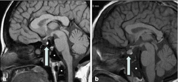
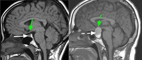
Normal vs abnormal pituitary mri. A Magnetic resonance image MRI and B corresponding schematic illustration of the human hypothalamus H and pituitary gland seen in saggital orientation. In the absence of the pituitary or of thyrotroph function hypothyroidism ensues. MRI is a powerful diagnostic imaging tool for detecting signs of injury like. The activity of the thyroid gland is predominantly regulated by the concentration of the pituitary glycoprotein hormone thyroid-stimulating hormone TSH. An MRI brain scan also shows brain lesions. 40 to 60 years Incidence.
Low levels can mean that your thyroid pituitary. Most people tend to blow off their Free T4 level because it isnt the free and active hormone especially when compared to free T3. By looking at MRI images your doctor can see details of blood flow and fluids surrounding the brain which can help determine abnormalities in the brain relating to arteries and veins. In addition in most instances MRI is the most sensitive imaging test and provides the highest quality images. Not all pituitary tumors called pituitary adenomas cause symptoms. Most patients do not require transsphenoidal hypophysectomy so diagnosis is based on history physical diagnostic imaging and the temporal relationship to pregnancy.
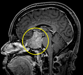
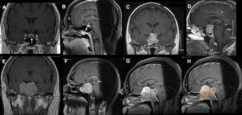
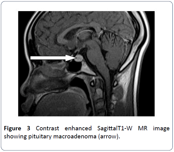
What is a Renin Test The renin test measures the amount of renin in the blood. Epidemiology Male Female. Magnetic resonance imaging MRI scan of pituitary macroadenoma. A prolactin test measures the amount of a hormone called prolactin you have in your blood. X-ray imaging tests including the pros and cons of each test how. They range between 4-23 ngmL mcgL in adult nonpregnant women and 3-15 ngmL in men 7 3.
The tissue thickens sheds and bleeds during every. Normal prolactin blood levels vary between sexes. The hormone usually rises if a woman is pregnant or has just given birth to help produce breast milk. Magnetic resonance imaging MRI is a noninvasive test doctors use to diagnose medical conditions. Blood is drawn from a vein usually from the inside of the elbow or the back of the hand. Clinical Radiology is published by Elsevier on behalf of The Royal College of RadiologistsClinical Radiology is an International Journal bringing you original research editorials and review articles on all aspects of diagnostic imaging including.
Brain lesions may be. A cerebral microbleed MB is a small chronic brain hemorrhage that likely results from structural abnormalities of your brains small vesselsDoctors can detect MBs with MRI sequences. Epidemiology Male Female. Magnetic resonance imaging MRI is an imaging technique used to evaluate many health conditions. Hypotonia is a lack of resistance to passive movement. Computed tomography Magnetic resonance imaging Ultrasonography Digital radiology Interventional radiology.
A brain lesion appears as a dark or light spot that does not look like normal brain tissues. Detailed MR images allow doctors to examine the body and detect disease. The puncture site is cleaned with antiseptic. Learn the ins and outs of MRI vs. 16-2 times Onset age. Quantitative parameters of magnetic resonance imaging cannot predict human epidermal growth factor receptor 2 HER2 status in rectal cancer.
TNF-α antagonists anecdotal reports Genetics Higher frequency of homozygous SMN2 deletion 40 than controls 10 17. Hypotonia is not a specific medical disorder but a potential manifestation of many different diseases and disorders that affect motor nerve control by the brain or muscle strength. View Media Gallery Hypothalamicpituitary surgery. MRI is unique to other imaging systems such as X-ray or CT scans because it does not require any harmful ionizing radiation. This International journal Journal of Clinical Neuroscience publishes articles on clinical neurosurgery and neurology and the related neurosciences such as neuro-pathology neuro-radiology neuro-ophthalmology and neuro-physiology. Sometimes healthcare providers order an MRI with contrast to help improve the diagnostic quality of the images.
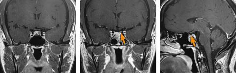
If a pituitary mass is suspected MRI is the best initial imaging study7 MRI is 61 to 72 sensitive and 88 to 90 specific for sellar. MRI with contrast can generate highly detailed visuals of internal organs and soft tissues. Identification of the disorder becomes clearer as the pituitary reverts to its normal size and recovers some of its normal function. Functional adenomas can cause problems because of the hormones they release. Normal range by age in years BUN. In healthy normal individuals the hypothalamus just above the brainstem sends CRH or corticotrophin-releasing hormone to the pituitary gland located behind the nose.
Being imaged immediately after TBI can result in more. The journal has a broad International perspective and emphasises the advances occurring in Asia the Pacific Rim region Europe. There are two major types of MRI scans. MRI does not use radiation x-rays. Adenomyosis is a medical condition characterized by the growth of cells that build up the inside of the uterus endometrium atypically located within the cells that put up the uterine wall as a result thickening of the uterus occurs. Hypotonia is a state of low muscle tone the amount of tension or resistance to stretch in a muscle often involving reduced muscle strength.
A magnetic resonance imaging MRI scan may be ordered to look for evidence of a pituitary growth or for damage to surrounding tissues. The first signs of a pituitary adenoma often depend on whether the tumor is functional making excess hormones or non-functional not making excess hormones. If sufficient normal tissue is excised inadvertently symptomatic hypogonadism may ensue initially followed by dysfunction of other pituitary cells. Thus regulation of thyroid function in normal individuals is to a large extent determined by the factors which regulate the synthesis. Chances are VERY high that your Free T4 level isnt actually normal even though it may be in the normal range. Blood tests to evaluate other hormone levels may be useful in ruling out any other possible causes of the symptoms.
An elastic band is placed around the upper arm to apply pressure and cause the vein to swell with blood. Diagnosis requires histopathologic examination. Note the high intensity or bright spot of the posterior pituitary by MRI in A sharply defining the boundary between the anterior pituitary gland. In case of abnormal results you may need to repeat the test to get the most accurate readings 2 3. This triggers the pituitary glands hyperproduction of adrenocorticotropic hormone ACTH causing the adrenal glands to flood the bloodstream with cortisol. As well as being misplaced in patients with this condition endometrial tissue is completely functional.
Incidence of pulmonary embolism and impact on mortality in patients with malignant melanoma.

T1 Weighted Mri Of Her Pituitary With Contrast Showing Normal Download Scientific Diagram
Iron And The Pituitary Gland Thalassemia Com
Cushing S Disease Ucla Pituitary Tumor Program


Imaging Of The Pituitary Gland Barrow Neurological Institute
Pituitary Tumor Symptoms Eyes Pituitary Tumor Boston Neec

Imaging Of The Pituitary Gland Barrow Neurological Institute
Finding The Right Solutions For Pituitary Tumors California Center For Pituitary Disorders

Surgical Treatment Of Pituitary Adenomas Endotext Ncbi Bookshelf

Conditions And Treatments Nasal Skull Base Pituitary Tumors Henry Ford Health System Detroit Mi
Surgical Treatment Of Pituitary Adenomas Endotext Ncbi Bookshelf
Pituitary Tumor North American Neuro Ophthalmology Society
Pituitary Adenoma Multiple Sclerosis And Visual Impairment How Are They Related A Case Report Insight Medical Publishing
View Of A Trial Of Oral Glucocorticoids In The Resolution Of Recurrent Granulomatous Hypophysitis A Case Report Journal Of The Asean Federation Of Endocrine Societies
![]()
Imaging Of The Pituitary Gland Barrow Neurological Institute
Posting Komentar untuk "Normal Vs Abnormal Pituitary Mri"