Syringomyelia Spinal Cord Cross Section
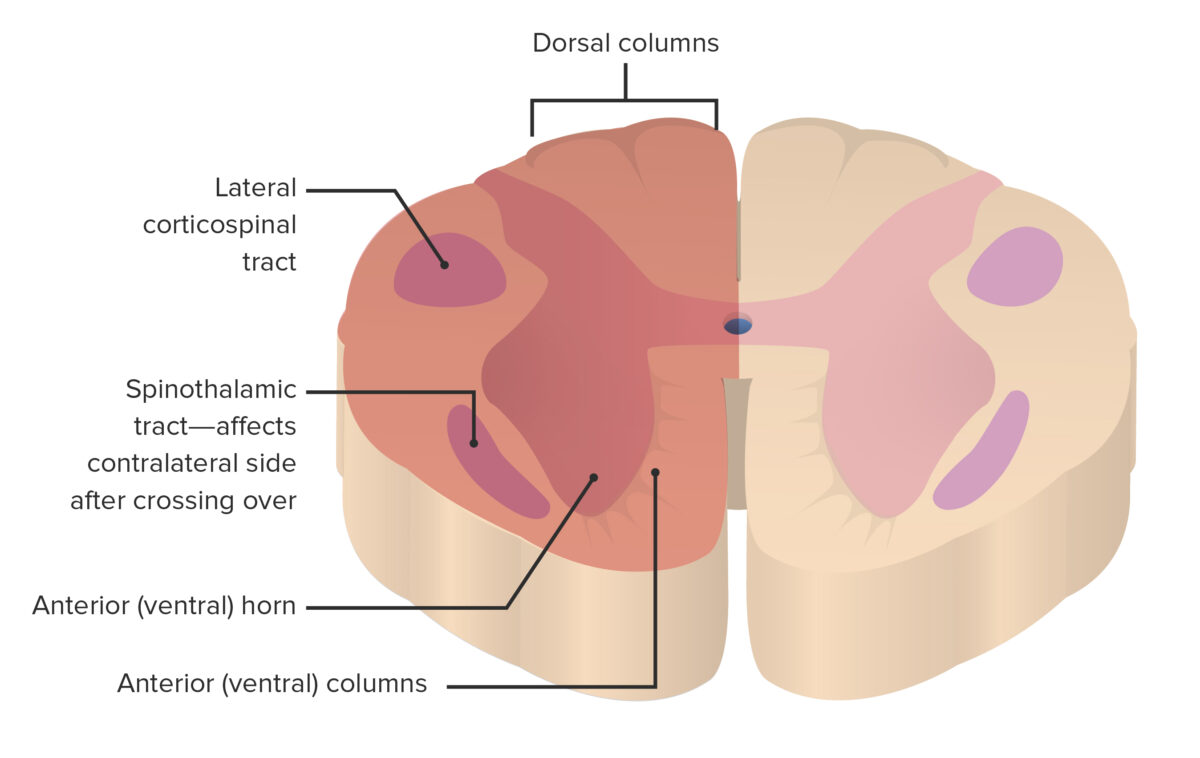
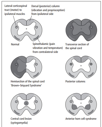
A lesion affecting approximately the left or right half of the spinal cord cross- section at one level creates a hemisection or Brown-Sequard syndrome Fig. There is cavitation within the substance of the spinal cord.
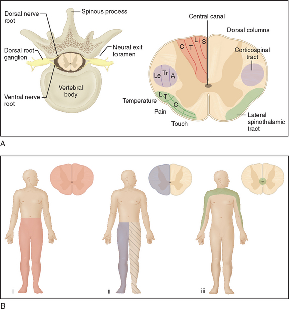
Suspended Sensory Level Neuroanatomy Flashcards Draw It To Know It
Many dogs with CM develop syringomyelia SM.

Syringomyelia spinal cord cross section. The syrinx may be asymptomatic being discovered incidentally on spinal cord imaging. Syringomyelia is a condition caused by a fluid-filled cavity or syrinx which forms within the spinal cord. Other symptoms you may have include. There was 1-2 of vacuole area per section in the spinal cord of laminectomy animals at 24 hours and 6 weeks Figure 7c. Syringomyelia is a condition in which an abnormal fluid-filled cavity or syrinx develops within the central canal of the spinal cord. A cross-sectional view of the spinal cord demonstrates a central butterfly-shaped area of gray matter and peripheral white matter Fig.
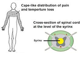
Pain progressive weakness in the arms and legs stiffness in the back shoulders neck arms or legs headaches loss of sensitivity to pain or hot and cold especially in the hands numbness or tingling imbalance loss of bowel and bladder control problems with sexual function. Syringomyelia is a disease which causes a softening or cavitation of the spinal cord anterior white commissure. Syringomyelia refers to cystic dilatation in the spinal cord usually secondary to various diseases such as Chiari malformation spinal cord injury tumor spinal arachnoiditis tethered cord etc 1234. As time goes on the syrinx enlarges damaging the spinal cord and causing a number of symptoms including. Syringomyelia is widely believed to be an acquired condition that is often idiopathic but can be seen following trauma spinal cord infarction cord infection noninfectious myelitis arachnoiditis or scoliosis or can be associated with intramedullary tumors such as hemangioblastoma or ependymoma Figs. METHODS All patients with a syrinx were identified from 14118 patients undergoing brain or cervical spine imaging at.
Spinal cord is located inside the vertebral canal. Despite extensive research the exact pathophysiology of cavity formation remains elusive and from a clinical perspec-tive syringomyelia remains a complicated condition to treat. A spinal segment is defined by dorsal roots entering and ventral roots exiting the cord ie a spinal cord section that gives rise to one spinal nerve is considered as a segment Figure 34. 76-5 and 76-6 In these cases the syrinx may. Syrinx of the Spinal Cord or Brain Stem. Each spinal nerve is composed of nerve fibers that are related to the region of the muscles and skin that develops from one body somite segment.
The peripheral white matter contains the axon tracts. Ad Offering a Full Range of the Latest Treatments for Syringomyelia. When the lower lobe of the brain the cerebellum is displaced to the level of the foramen magnum mild CM or through the foramen magnum severe CM there is overcrowding in the foramen magnum. OBJECT Syrinx size and location within the spinal cord may differ based on etiology or associated conditions of the brain and spine. Curving of the spine called scoliosis Changes in or loss of bowel and bladder function Heavy sweating Not being able to feel hot and cold in the fingers hands arms and upper chest Loss of reflexes Muscle stiffness that may make it. Typically the lesion is around and most often anterior to the central canal.
Predisposing factors include craniocervical junction abnormalities previous spinal cord trauma and spinal cord tumors. A syrinx can refer to an abnormal gliotic-lined fluid-filled cavity located within the spinal cord parenchyma syringomyelia or a focal dilation of the central canal hydromyelia. These differences have not been clearly defined. Ventral abdomenthroat Dorsal back Ventral abdomenthroat Dorsal back Ventral Horn of Grey Matter. This causes obstruction of the normal flow of CSF from the brain down to the spinal cord. When a person develops syringomyelia they develop a syrinx a cyst filled with fluid within the spinal cord.
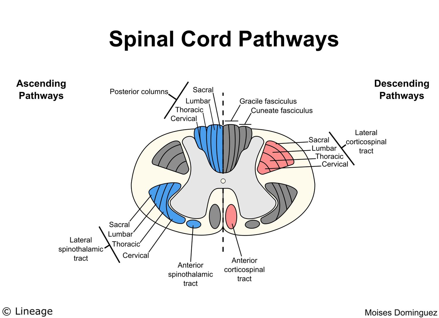
In cross section the spinal cord is divided into an H-shaped area of gray matter consisting of synapsing neuronal cell bodies and a surrounding area of white matter consisting of ascending and descending tracts of myelinated axons. Reveal how Syringomyelia develops and common causes to know about now. Average age at onset is 30 years. Symptoms usually appear late in the second decade through the fifth. Transverse cross-section of the spinal cord butterfly in centre is grey matter. 12 These two terms have been used interchangeably in the literature and.
The dilation of the cord leads to abnormalities in the neurologic pathways that transmit pain and temperature. The syrinx is a result of disrupted CSF drainage from the central canal commonly caused by a Chiari malformation or previous trauma to the cervical or thoracic spine. Up to 10 cash back A cross section of the spinal cord SC reveals the white matter consisting of ascending and descending tracts arranged around the gray matter consisting of neuronal bodies unmyelinated motor-neuron fibers and interneurons. The percentage of vacuole area per spinal cord cross section became obvious at 24 hours onwards in laminectomy and syrinx groups. This percentage increased by 36- and 61-fold in syrinx at 24 hours and 6 weeks respectively. Doctors Work with You to Choose the Treatment that Best Suits Your Needs.
Syringomyelia is a rare spinal cord condition characterised by the formation of large uid- lled cavities in the spinal cord. End of the spinal cord are between L1 and L2. Syringomyelia produces bilateral deficits in pain and temperature in the body areas innervated by the affected cord segments. Symptoms include flaccid weakness of the hands and arms and deficits in pain and. Using a plated bayonet the cord is bluntly split open until the syrinx is reached and opened Fig. Syringomyelia is rare with a prevalence of 8 per 100000 persons.
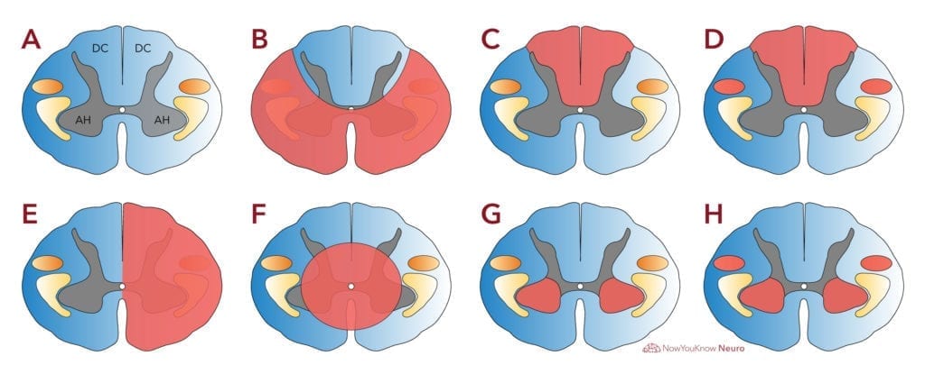
Syringomyelia is defined as cystic dilation of the central canal of the spinal cord. Ad Discover why Syringomyelia symptoms is not too complicated to spot right now. A syrinx is a fluid-filled cavity within the spinal cord syringomyelia or brain stem syringobulbia. Syrinx affecting BOTH dorsal and ventral horns yellow green pain movement affected. The central gray matter contains the neural cell bodies. Involvement of the spinothalamic tract produces a contralateral deficit to pain and.

Simplistic Representation And Corresponding Mr Images T1 Of The Csf System Around The Brain And Cervical Spine For A Chiari Patient With Syringomyelia Showing Location Of The Syrinx Blue Region In Center
Diffusion Weighted Mr Imaging In A Rat Model Of Syringomyelia After Excitotoxic Spinal Cord Injury American Journal Of Neuroradiology
Spinal Disease Neoplastic Degenerative And Infective Spinal Cord Diseases And Spinal Cord Compression Neupsy Key

Diseases Of The Spinal Cord Fundamentals Of Neurology An Illustrated Guide
Brown Sequard Syndrome Concise Medical Knowledge
Spinal Cord And Spine Anatomy Review Chapter And Practice Questions
Spinal Cord Lesions Disorders Of The Spinal Cord
Numbness Of The Hands Legacy Spine Neurological Specialists
Spinal Cord Neurology Medbullets Step 1

Chiari And Syringomyelia 101 Page 3 Asap American Syringomyelia Chiari Alliance Project

Figure 2 From Anomalous Development Of The Spinal Cord In A Calf Semantic Scholar

Figure 2 From Clinical Reasoning A 39 Year Old Man With Abdominal Cramps Semantic Scholar

Cross Sections Of The Pressure For Ascending A D And Descending E Download Scientific Diagram
Posting Komentar untuk "Syringomyelia Spinal Cord Cross Section"