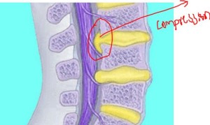
Thecal Sac Compression C3 4
Thecal sac and cord noted. The short answer is YES.
Disc Bulge Disc Bulge On Thecal Sac

MRI white matter lesions Many times I get consulted by patients or their relatives when their MRI brain report reads multiple scattered white matter lesions seen.

Thecal sac compression c3 4. Only original papers are considered for publication with the understanding that they are contributed solely to Spine. The mean pre- and post-operative MAS and PROM were 28 and 04 p 0001 395o and 660o p 0001 respectively. C5- C6 grade 1 retrolisthesis. Diagnosing brachial plexus pathology can be clinically challenging often necessitating further evaluation with MRI. Patients and caregivers are naturally worried when. Facet joint injections - An initial facet injection intra-articular and medial branch block from C2-3 to L5-S1 is considered medically.
Facet joints are unremarkable. It does appear to be foraminal narrowing seen at C7-T1 bilaterally. Its quite an experience hearing the sound of your voice carrying out to a over 100 first year. Mild moderate right neural foraminal narrowing. Minimal if any neural foraminal. There is desiccation of the T10 T11-T12 disc.
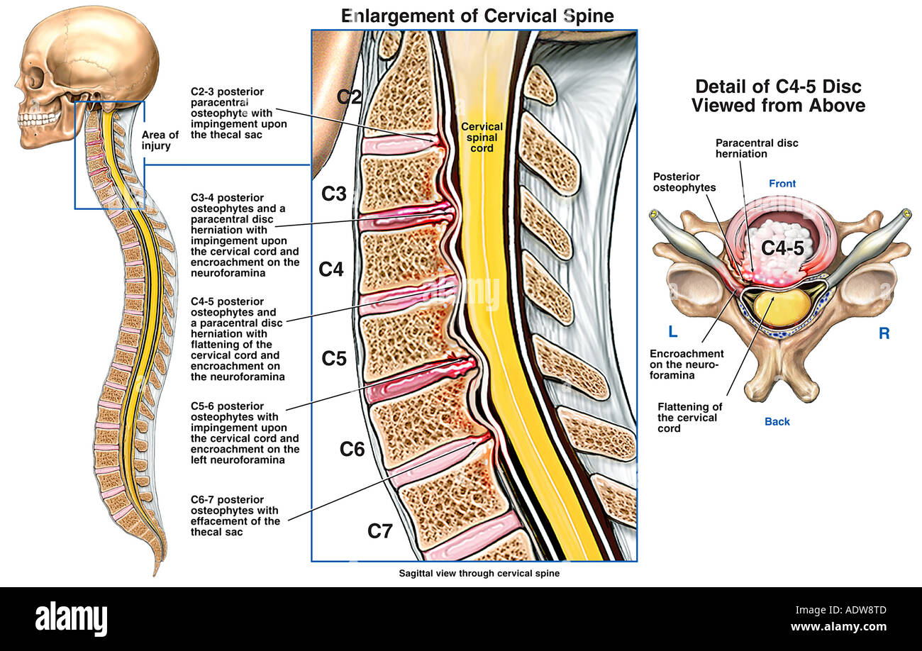
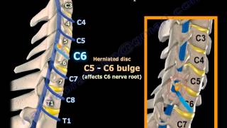
It is the leading subspecialty journal for the treatment of spinal disorders. L4-L5 - level is desiccated and narrow. Spine is an international peer-reviewed bi-weekly periodical that considers for publication original articles in the field of spine. It showed a C5-C6 paracentral disk protrusion moderate in size compressing upon the thecal sac. Epidural fat surrounds the thecal sac and is less dense than soft tissue so it appears darker gray. B Axial T2-weighted MR image shows the mass white arrow occupying the left half of the thecal sac displacing the spinal cord to the right with an adjacent CSF flow void arrowhead in the right posterolateral subarachnoid space.
Rotation as high as an average 79 was seen in one study of adult volunteers and loss of contact of the articular facets of C1 and C2 during rotation as high as 74 85 seen in physiologic conditions was. The microscope was brought in direct with microdissection there was a massive disk herniation on the right side underneath the nerve root as well as the left. Some narrowing of the lateral recess noted bilaterally. Erected vertically the spine is the mast of our body and has three major functions. Is a minimal bulge without evidence of a focal protrusion again no significant encroachment seen of the central canal or foramina. Aetna considers any of the following injections or procedures medically necessary for the treatment of back pain.
Academiaedu is a platform for academics to share research papers. Paraspinal soft tissues appear unremarkable. C3-4 mild disc bulge with narrowing of central canal. This difference in tissue density allows discrimination of the outer margins of the thecal sac. C Axial T2-weighted image shows the ventral epidural high T2 collection indenting the ventral thecal sac and abutting the cord. L5-S1 - level is desiccated.
The thecal sac was retracted medially. Significant impression upon the thecal sac. A Google ingyenes szolgáltatása azonnal lefordítja a szavakat kifejezéseket és weboldalakat a magyar és több mint 100 további nyelv kombinációjában. Chiropractic Specialty Center uses specialized devices methods and care systems that are significantly superior to the traditional techniques of non-invasive. B Sagittal T1 postcontrast image shows large ventral collection with peripheral enhancement causing mass effect upon the cord. A Sagittal view of a large paracentralforaminal disk protrusion at the C4-C5 level indenting the anterior thecal sac and flattening the anterior surface.
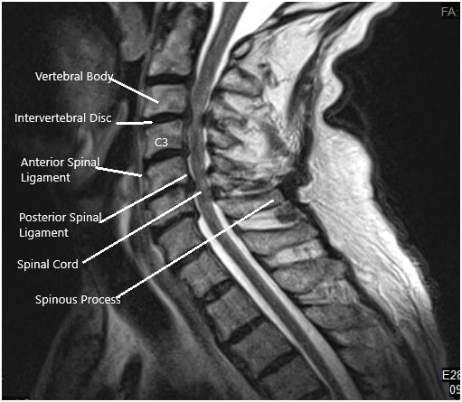
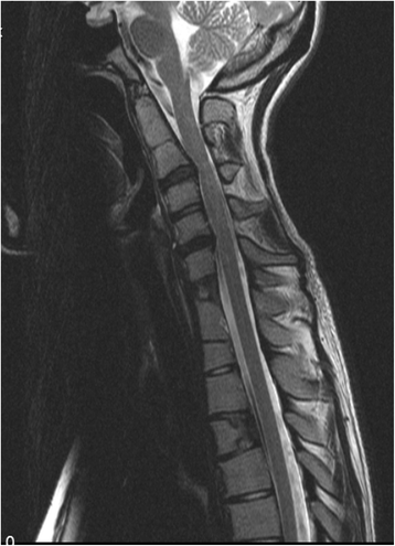
Note discitis at the C3-C4 level. A total of 21 STNs were performed in 15 patients. He reports he dove into a lake without understanding the depth and hit a rock. Owing to its vague symptomatology uncommon nature and complex anatomy the brachial plexus presents a diagnostic dilemma to clinicians and radiologists alike and has been the subject of many prior reviews offering various perspectives on its imaging. OBQ1783 A 21-year old previously healthy male presents to the trauma bay 8 hours after a helicopter evacuation from a national park with a suspected cervical spine injury. Transverse T2W image showing the C3-4 level with no spinal cord compression.
The Journal does not publish articles reporting material that has. He treated for four months from the date of the accident. Does thecal sac compression or dura sac compression mean spinal cord compression or nerve root compression. Moderate severe left neural foraminal narrowing. What is the code assignment for stenosis of the cervical spine at C3-C6 with myelopathy. Mild moderate broad-based disc stripe complex effacement of the thecal sac and compression of the spinal cord.
Moderate narrowing of the foramen bilaterally. Epidural abscess not well seen on CT. Disc bulge is seen. The radiologists report usually further reads that these can be seen in primary demyelinating conditions like multiple sclerosis or in vascular disorders. About 1 month later he had an MRI of the cervical spine. Provided however that only one invasive modality or procedure will be considered medically necessary at a time.
At the end of his treatment the diagnosis was a lumbar strain. The mean level of MGHFAC was improved from 33 pre-operatively to 49 post-operatively p 001. C Axial CT image shows dense ossification of the mass arrow correlating with the very low T2 signal intensity. Enter the email address you signed up with and well email you a reset link. Notes that had issues from Thoraci MRI T8-T9 demonstrates a mild left paracentral protruded disc with minimal effacement of ventral thecal sac. To provide structural support enable trunk movement and protect the neural elements From a biomechanical point of view the spine is a multiarticular structure comprising numerous segments or units enabling multidirectional motions and the absorption of large.
6 non-ambulators had significant amelioration in MGHFAC level. The AP thecal sac is estimated at 7-8 mm. Facet ligament overgrowth results in a trefoil appearance the thecal sac and mild narrowing of the canal. Conservative treatment of ligamentum flavum hypertrophy is effective when provided by a team with an expert understanding of the spine and associated conditions. Narrowing of the neural foramina greater on the right than the left. On primary exam he has a 5 cm laceration over the parietal region of his skull with no other aparent injuries in his extremities or.
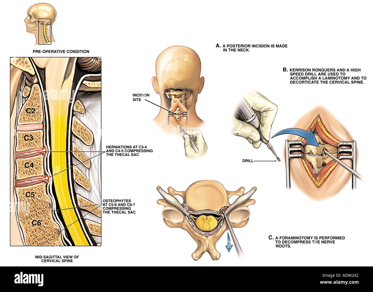
Ing in compression of the thecal sac and its contents. The disc was incised with an 11-blade knife and was cleaned out with a series of straight and angled curettes and rongeurs.
Case Study Management Of 73 Year Old Female With Cervical Spine Stenosis And Myelopathy

Medpix Case Disc Herniation With Resultant Spinal Cord Injury

2 Level C3 4 And C5 6 Cervical Spine Disc Herniation Medical Art Works
Severe Thecal Sac Compression Back 17 Questions Answered Lybrate Ask Questions
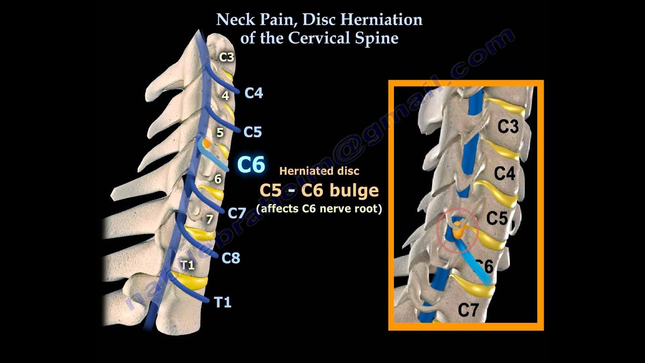
Neck Pain Disc Herniation Of The Cervical Spine Everything You Need To Know Dr Nabil Ebraheim Youtube
Cervical Vertebrae C7 High Resolution Stock Photography And Images Alamy
Thecal Sac Compression Effacement What Is Indentation
Degenerative Disc Neck High Resolution Stock Photography And Images Alamy
Neck Pain Disc Herniation Of The Cervical Spine Everything You Need To Know Dr Nabil Ebraheim Youtube

Plain Radiograph Computed Tomographic Scan And Magnetic Resonance Download Scientific Diagram
Cervical Instability In Klippel Feil Syndrome Case Report And Review Of The Literature Chinese Neurosurgical Journal Full Text
A Patient Presenting With Csm With Predominantly C5 C6 And C6 C7 Download Scientific Diagram

Medpix Case Disc Herniation Complex At C3 4 And 5 6 Disc Bulge At C4 5 And C6 7facet Joint Hypertrophy With Neuroforaminal Stenosis Bilateral At L5 S1high Grade Tear In Supraspinatus Tendon
Diffuse Cervical Bulge Orange County Disc Associates

Posting Komentar untuk "Thecal Sac Compression C3 4"