Left Middle Cranial Fossa Arachnoid Cyst
Symptoms vary by size and location. A fossa cyst is a liquid-filled cyst which forms in a fossa area in the body.
22 Year Old Man With Complicated Arachnoid Cyst Postoperative Download Scientific Diagram

No evidence of acute ischemia.

Left middle cranial fossa arachnoid cyst. We here report the unusual case of a young patient with an arachnoid cyst. In one reported study 18 of middle cranial fossa cysts presented with progressive symptoms whereas 44 presented with nonprogressive symptoms and 37. Arachnoid cysts of the posterior fossa are rare. Arachnoid cysts are congenital extra-axial non-neoplastic lesions filled with CSF 12 which is a common incidental discovery on neuroimaging in all age groups 2. Arachnoid cysts involving the spinal cord are rarer. This could be confirmed on follow up MRI.
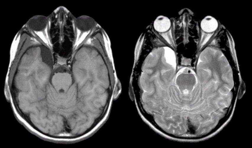
While arachnoid cysts vary in their location most are supratentorial and found in the middle fossa. The location and size of the cyst determine the. A Sagittal T1-weighted image 65020 dem onstrates hypogenesis of left middle temporal gyrus arrow. Providers call this membrane the arachnoid membrane because it looks like a spider web. They are benign in nature and congenital in origin. The majority of arachnoid cysts form outside the temporal lobe of the brain in an area of the skull known as the middle cranial fossa.
B Axial T2-weighted image 300090. The majority of arachnoid cysts form outside the temporal lobe of the brain in an area of the skull known as the middle cranial fossa. Arachnoid cysts are benign and the vast majority remain asymptomatic throughout life 2. Neuropsychological examinations Digit Span. Of AC in the left middle cranial fossa and left orbital meningocele with communication between the two cysts through a small bony defect in the left lateral wall of orbit was given. Margin of temporal lobe adjacent to middle cranial fossa arachnoid cyst is undulating arrow and there.
The majority of arachnoid cysts form outside the temporal lobe of the brain in an area of the skull known as the middle cranial fossa. Non-contrast enhanced computed tomography CT scan of orbits was performed to. Arachnoid cysts are intra-arachnoidal space-occupying lesions containing fluid similar to CSF. Three cases are presented with a previously unreported otologic symptom that of bilateral hearing loss which i. Arachnoid cysts represent 1 of all nontraumatic intracranial masses. When arachnoid cysts are encountered the presenting symptoms are frequently otologic with hearing loss and imbalance occurring commonly.
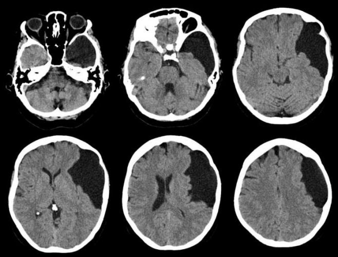
The location and size of the cyst determine the symptoms and when those symptoms begin. However the cranial CT scans of both patients revealed small arachnoid cysts in the left middle cranial fossa extending into the adjacent sylvian fissure without a marked mass effect on either the temporal or frontal lobes and the lateral ventricle Figure 1. Arachnoid cysts are developmental collections of cerebrospinal fluid covered by layers of arachnoidal epithelium and are usually located in the middle cranial fossa. Arachnoid cysts involving the spinal cord are rarer. Headaches are the most common symptom accounting for a share of 66 6. We discuss the relationships of aneurysm arachnoid cyst and subdural hematoma.
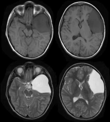
The Galassi classification of middle cranial fossa arachnoid cysts uses the size and degree of displacement of the adjacent. An EEG revealed no abnormality. Both types develop on a thin membrane that covers and protects the brain and spinal cord. Intracranial arachnoid cysts usually occur adjacent to arachnoidal cistern and may present with hydrocephalus. There is a fluid density collection in the left middle cranial fossa 35 x 47 cm with an imperceptible wall and thinning of the overlying bone in keeping with an arachnoid cyst. What is an arachnoid cyst.
Localizations in the posterior fossa are uncommon and generally remain asymptomatic or cause vague and non-specific symptoms. We report a case of a cerebral aneurysm arising from the bifurcation of the left middle cerebral artery that ruptured into a left middle cranial fossa arachnoid cyst associated with acute subdural hematoma. 66 23 of intracranial arachnoid cysts occur in the middle fossa. Intracranial or spinal cavities containing a cerebrospinal-like fluid the wall of which is composed of arachnoidal cells. We describe a rare presentation of an arachnoid cyst which ruptured after a minor head injury and resulted in a subdural hygroma. The most common location for this entity is the middle cranial fossa 12.
A cyst is sac-like growth which forms in the body and is filled with fluid gas or a solid matter. Diagnosis and treatment of arachnoid cysts of the posterior. Small cysts usually have no symptoms and are discovered only incidentally. They are most often developmental or related to trauma. The location and size of the cyst determine the symptoms and when those symptoms begin. It exerts mass effect on the left temporal lobe and left lateral ventricle.
And focal neurologic signs. Arachnoid cyst is cerebrospinal fluid CSF filled sac not tumors not cancer that is located on the arachnoid membrane that covers the brain intracranial and the spinal cord spinal one of the three membranes that cover the brain and spinal cord 1. 2-33-year-old woman with left middle cranial fossa arachnoid cyst. These usually begin while the brain and skull are forming in the womb. The cysts can form in several areas of the brain. Ad Learn the early warning signs and causes of arachnoiditis to be aware of today.
The most troublesome type of cyst is located in the brain also known as an arachnoid fossa cyst or cranial cyst. Arachnoid cysts involving the spinal cord are rarer. Most arachnoid cysts grow in the middle fossa region located in front of the ears. Other symptoms include dizziness nausea vomiting worsening of mood mental status changes ataxia seizures and hearing loss 7.
Cystma
Cystmnn2
Intracystic Hemorrhage Of The Middle Fossa Arachnoid Cyst And Subdural Hematoma Caused By Ruptured Middle Cerebral Artery Aneurysm American Journal Of Neuroradiology

Update Sylvian Fissure Arachnoid Cyst Neurosurgery

An Aneurysm Rupturing Into A Middle Cranial Fossa Arachnoid Cyst Presenting As An Intracystic Hemorrhage Journal Of Stroke And Cerebrovascular Diseases
Mri And Ct Scans Of The Brain Showing Arachnoid Cyst In The Left Middle Download Scientific Diagram

Intracranial Arachnoid Cyst Operative Neurosurgery

A Axial Ct Showing An Arachnoid Cyst In The Left Middle Cranial Fossa Download Scientific Diagram
Middle Fossa Arachnoid Cysts And Inner Ear Symptoms Are They Related Semantic Scholar

Cyst Post
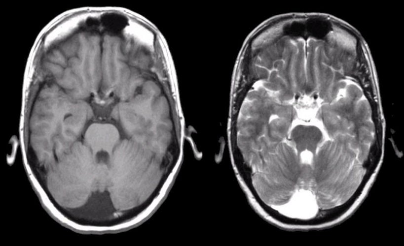
Figure 3 From Endoscopic Fenestration Of Middle Cranial Fossa Arachnoid Cysts Does Size Matter Semantic Scholar

Cyst

Cystma3
Hyperdense Extra Axial Cyst In The Right Anterior Middle Cranial Fossa Download Scientific Diagram


Posting Komentar untuk "Left Middle Cranial Fossa Arachnoid Cyst"