How To Read Visual Field Test Results
A test that shows visual field loss means that vision in some areas is not as sensitive as normal. GLAUCOMA HEMIFIELD TEST GHT The GHT provides a plain language classifi cation of 30-2 and 24-2 test results based upon patterns of loss commonly seen in glaucoma.

Understanding Glaucoma Test Results What Do They Mean
Look at the GHT mean deviation VFI and pattern standard deviation.
How to read visual field test results. The mean deviation represents the degree of departure of the whole fields average values from age-adjusted normal values. Points along circles known as isopters Figure 3. Look at the pattern. The Goldmann Visual field tests the entire visual field one eye at a time by plotting. The size and. Choplin NT Edwards RP.
Look at the reliability indices. Patients were classified as pass or fail by both the integrated visual field and the Esterman test. Interpreting the Goldmann field test. Patients performed the bilateral monocular field tests to generate the integrated visual field the Esterman test and the UFOV test on the same visit. Use this order to interpret your Humphrey visual field every time. Mean Deviation MD represents the average sensitivity deviation from a normal healthy person of the same age based on the normative data base.
It can be seen from this sample scan that there is a dark line which represents the RNFL for this patient and that there are quadrant areas where this line crosses into the outside normal limits area. Theyll ask you to look directly at them then theyll hold up their hand and move it back and forth. A Text and Clinical Atlas. Youll then signal when their hand appears in your vision. Look at the reliability indices. Factors influencing visual field measurements CLINICAL VARIABLES Pupil size It is best to test the visual field with a pupil diameter of at least 3 mm record pupil size when testing visual field Fixation Important to monitor during visual field testing Target blur Media opacities eg cataracts Age 14 decreased sensitivity.
The visual field index is a staging index designed to cor- respond to ganglion cell loss that is 100 represents normal fields and 0 represents blind fields. The skill is in identifying patterns and observing any change with repeated tests. Compare to the previous visual fields. It could be just a little vision lost in a small area or all vision lost in large areas. People also ask what do the numbers mean on a visual field test. By recognizing the strengths and limitations of visual field testing the practitioner can more accurately diagnose and follow patients with glaucoma.
While you continue to stare at the centre of the screen small flashes of light will briefly appear in your periphery and they. UFOV risk scores were calculated for each patient. Links to different parts of the new lecturePart 1. Confrontation Visual Field Exam. During a confrontation visual field exam your doctor will sit a short distance in front of you. Visual fields that are not reliable contain artifacts or cannot be trusted for other reasons should be retested if this is clinically relevant.
The MD is an indication of the overall VF sensitivity and can either be a negative or. The key to interpreting Goldmann visual fields is to keep in mind the normal hill of vision figure 1 and how it compares with the patients results. Other methods to test the visual function are for example contrast sensitivity test field of vision test and colour test charts. FDT provides two global indices to generally summarize the visual field results for threshold tests similar to HFA white-on-white perimetry 1. Doctors can use these results in association with the visual field test to determine how RNFL regions correlate with test scores of VF regions. Click to see full answer.
Firstly a visual field test is a simple and painless test which measures your peripheral or all-around vision. Confirm its the rightleft eye. Confirm its the right patient with name and date of birth. Visual Field Testing With the Humphrey Field Analyzer. Furthermore visual acuity does not tell you everything about the functioning of the eye. STEP 2 ASSESS WHETHER THE VISUAL FIELD CAN BE TRUSTED FIGURE 8-5 Before interpreting visual field results it is important to confirm that the visual field can be trusted.
65 patients were. During it an optical assistant will ask you to cover one eye place your chin on a rest and gaze at a central point of a screen which is usually white. Look at the GHT mean deviation VFI and pattern standard deviation. Look at the pattern. This will give the doctor a general idea of your peripheral vision so they may use it as an. Area tested - Stimulus si.
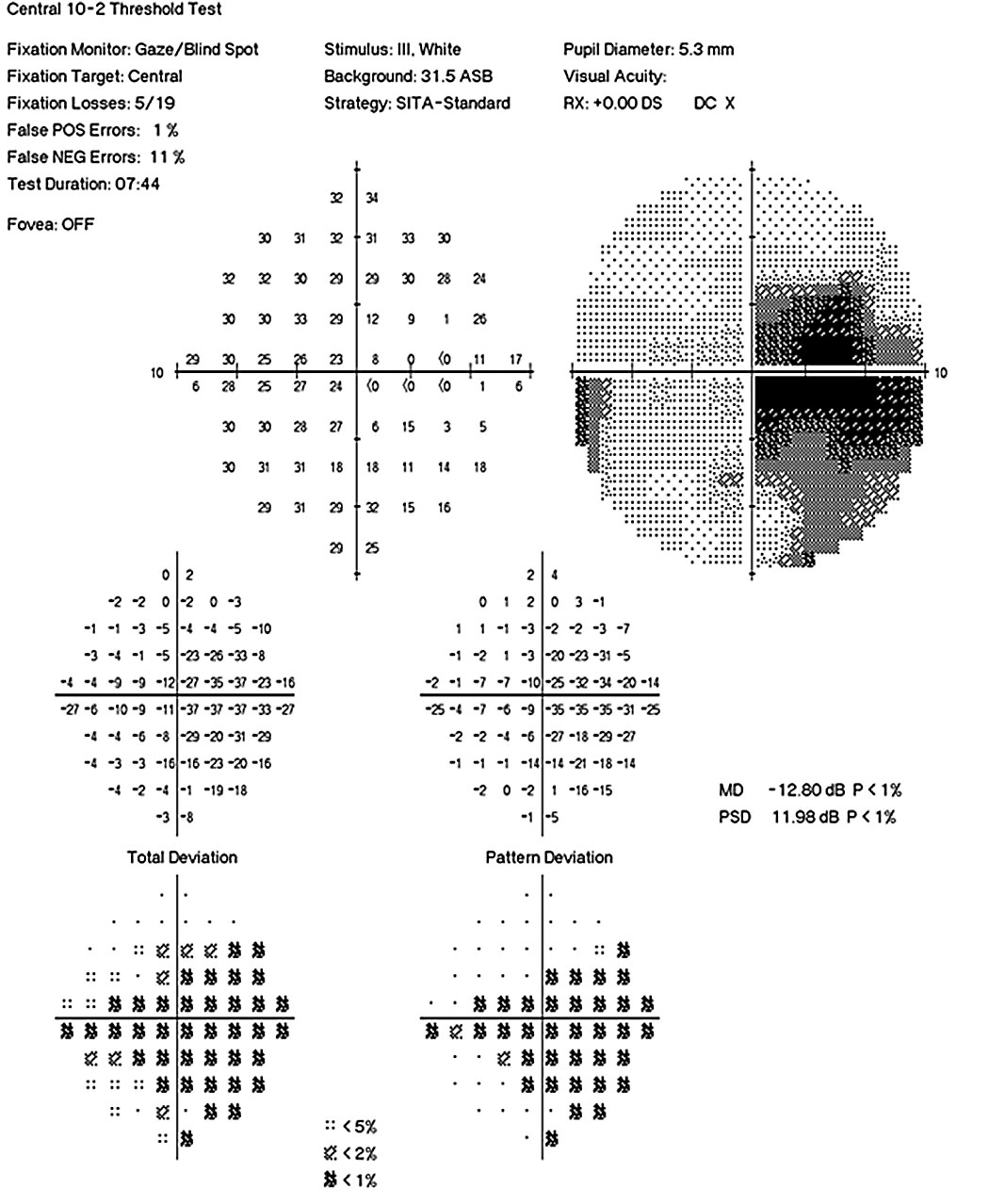
Pattern Deviation scores in each of fi ve zones in the upper hemifi eld are compared to fi ndings in mirror-image zones in the inferior visual fi eld. Mayer explains how to interpret automatic static perimetry resultsSpecial educators can earn professional credits for watching this video. The amount of vision lost and the areas affected are. Higher numbers mean the patient. Each isopter should be color-coded to. Confirm its the right patient with name and date of birth.
Confirm its the rightleft eye. A normal visual field test means that the patient can see about as well as anyone else does in the center and around the edges of the visual field.

Quadrantanopia An Overview Sciencedirect Topics
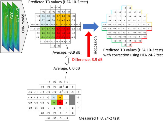
Predicting The Central 10 Degrees Visual Field In Glaucoma By Applying A Deep Learning Algorithm To Optical Coherence Tomography Images Scientific Reports
The Neurologic Exam Step By Step
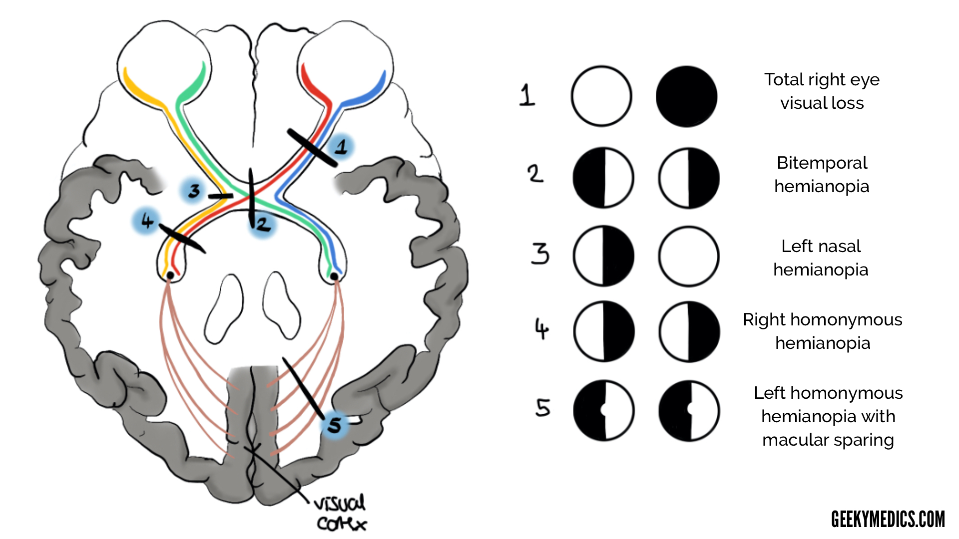
Visual Pathway And Visual Field Defects Geeky Medics
Visual Pathway And Visual Field Defects Geeky Medics

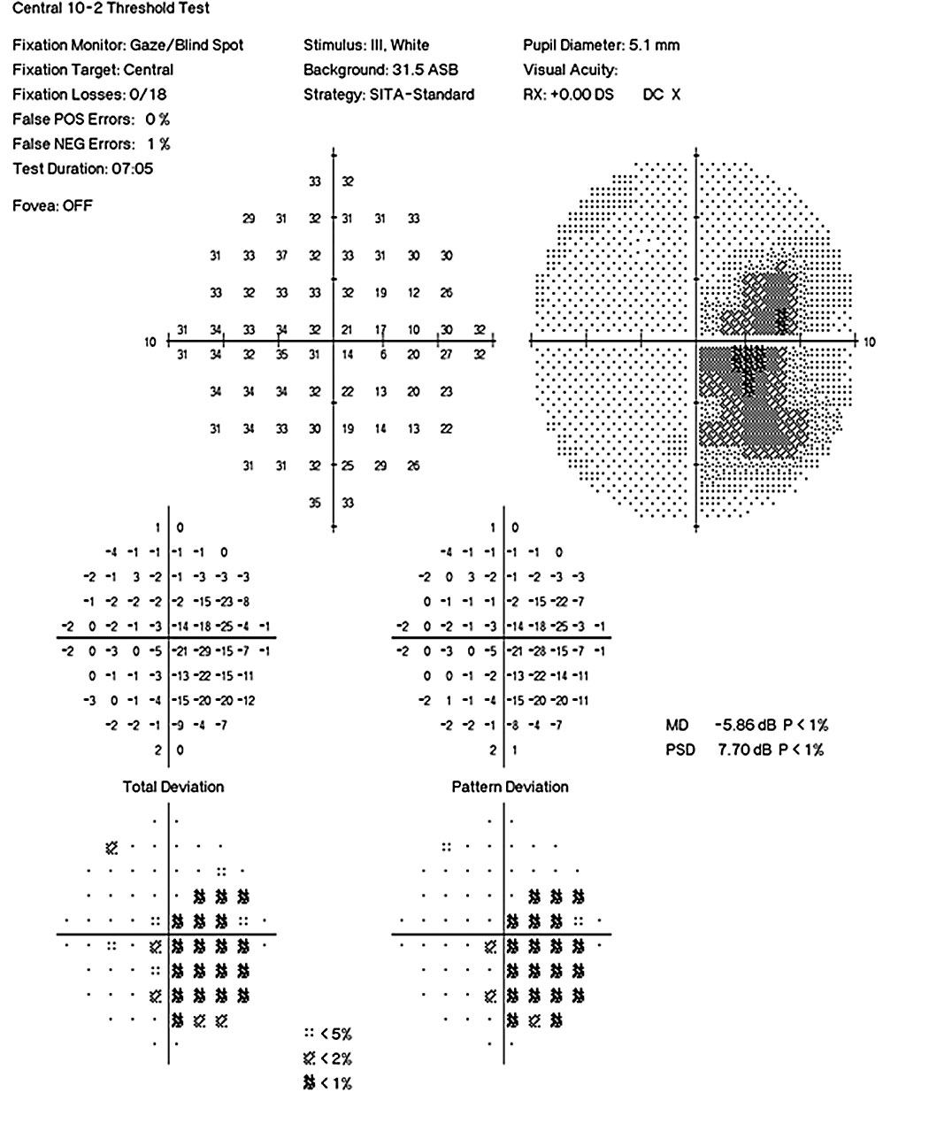
Simultaneously Performed Combined 24 2 And 10 2 Visual Field Tests In Glaucoma Scientific Reports

Typical Glaucomatous Visual Field Loss Both The Grayscale And Pattern Deviation Plots Are Shown Optometry Education Optometry Eye Health Facts
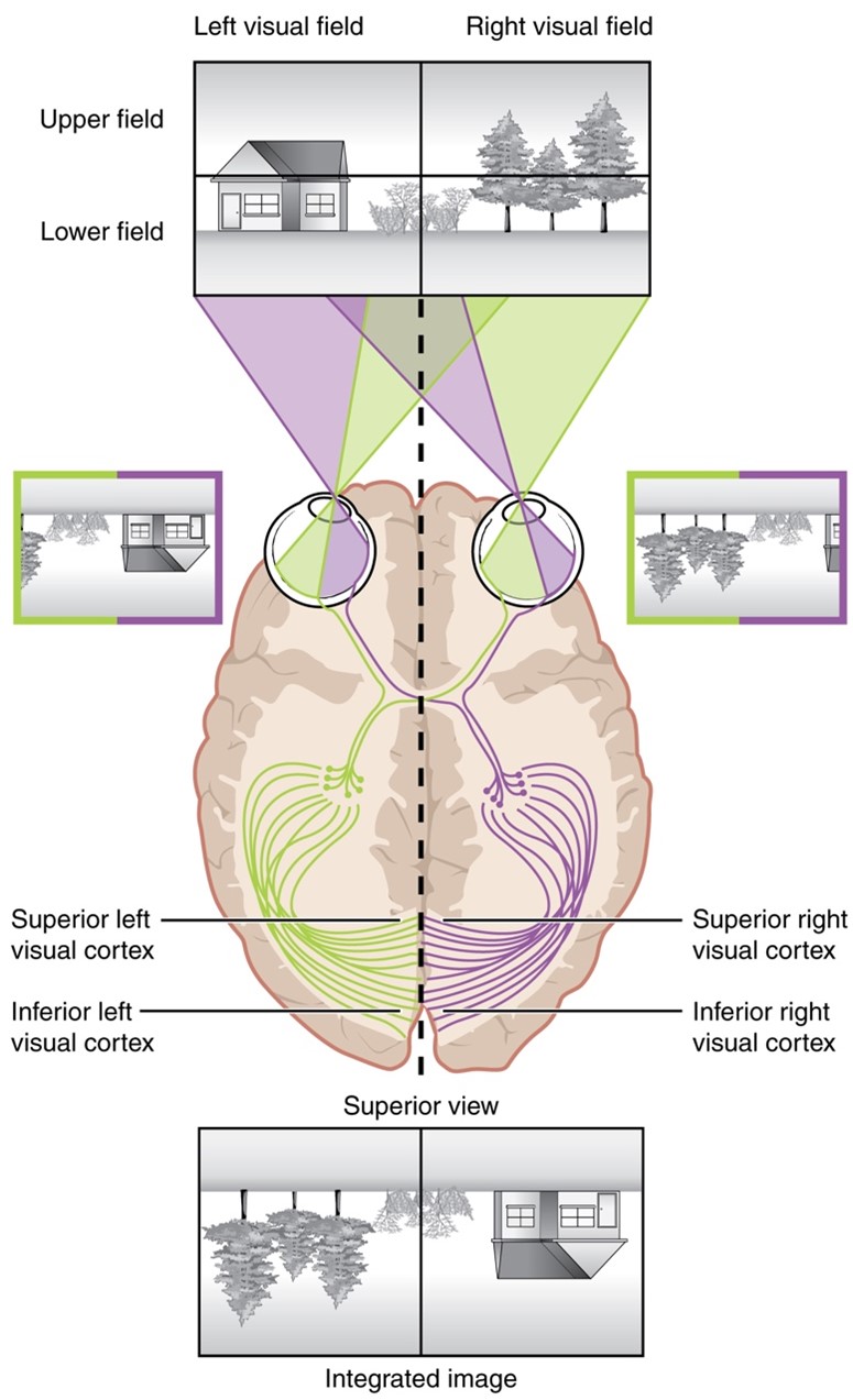
Neuroscience For Kids Visual Pathway

Examination Of Visual Field By Confrontation With The Examiner Approximately 18 Inches 45 Cm From The Patient The Patient S Unteste Visual Nclex Prep Nclex

Visual Field
2
The Neurologic Exam Step By Step

Visual Field

Visual Field
Posting Komentar untuk "How To Read Visual Field Test Results"