Mri Pictures Of Cervical Herniated Disc
A herniated disc may be caused by injury or degeneration from age. Injury an injury in your neck or spine region is an instant aggregator for cervical pain.
Cauda Equina Syndrome From Lumbar Disc Herniation Cauda Equina Cauda Equina Syndrome Lumbar Disc

Some hospitals may offer promos to their patients who may need to undergo more than one MRI scan.

Mri pictures of cervical herniated disc. How to Relieve Pins and Needles in Hands. The material inside of the disc can then spill out. Symptoms depend on the location of the herniation and whether nerve tissue is being irritated. Most people with low back pain do not require a CT or MRI. The MRI is the most commonly used test to evaluate the spine because it can show abnormal areas of the soft tissues around the spine. The cervical spine is the portion of your spine that runs through your neck.
Disc bulging disc herniations annular tears spinal stenosis neuroforaminal narrowing. 56 Richardson 11 also reports a case of NP with associated neck upper back and low back pain of approximately two years duration with symptoms that began following a rear-end motor. A common cause of pinched nerves in the back is a herniated disc. Radiating nerve pain or radiculopathy is a common symptomAs a small hard lump intrudes on the dense system of nerves concentrated within and around the spinal column pinched nerves can ensue. In fact a herniated disc is seen on CT scan or MRI in 25 percent of people without low back pain. The MRI is done to find tumors herniated discs or other soft-tissue disorders.
One of the common findings is a diffuse disc bulge at L4-L5. The MRI could lead to a diagnosis of infection tumor inflammation or pressure on your nerve. When the degeneration of the joint is secondary to natural wearing and abnormal body mechanics the condition is known as osteoarthritis OA. The MRI is better than X-ray because in addition to the bones it can also show pictures of the nerves and discs. This means that the area of the disc bulge is large and spread out. A magnetic resonance imaging MRI indicates a herniated disk at the L3-L4 level that is pressing on the right L4 nerve root of his spine.
My MRI or X-ray Says I Have a Loss of Cervical Lordosis or a Problem with My Intervertebral Discs. We are going to look at examples of each of these conditions. It is performed to remove the large arthritic bone spurs and a portion of the herniated discs compressing the spinal nerves. A herniated spinal disc or trapped nerve in the neck can apply pressure on a group of nerves that can cause numbness in the arms and hands. However before we do it is important that you understand that after the age of 30 essentially every MRI scan of the human body is abnormal. MRI does not use radiation x-rays.
Damage can occur as a result of pressure from material from a ruptured disc degenerative changes in bones arthritis or other injuries that put. Bulging discs or herniated discs. Every MRI scan after the age of 30 is abnormal. He received his MD from the Albert Einstein College of Medicine in 2010 followed by a residency at the Oregon Health Science University and fellowship at the University of California Davis. Also several injection techniques are helpful to learn the conditions causing hip pain. The MRIs revealed the following.
25 mm protrusion with annular tear. The MRI is the most commonly used test to evaluate the spine because it can show abnormal areas of the soft tissues around the spine. A herniated or ruptured disc is usually more serious than a bulging disc. However some patients may require additional diagnostic imaging such as X-ray magnetic resonance imaging MRI computerized tomography CT or ultrasonography. Toms physician offers a transforaminal lumbar epidural steroid injection TFESI as one strategy to. This occurs when the soft center of a disc known as the nucleus pushes through the harder outer disc layer called the annulus.
The outer protective layer of the disc remains intake with a bulging disc but cracks or ruptures with a herniation allowing some of the protective material inside to leak out. Bone fracture and injuries. A smaller disc bulge often means the bulge comes out further but it is less spread out while a diffuse disc bulge at L4-L5 means the opposite. The MRI is done to find tumors herniated discs or other soft-tissue disorders. The MRI is better than X-ray because in addition to the bones it can also show pictures of the nerves and discs. The MRI produces pictures of soft tissues as well such as ligaments tendons and blood vessels.
Herniated discs this kind of cervical pain occurs when spinal discs develop cracks in them. This article was medically reviewed by Troy A. However I have had three MRIs each with progressive findings. My entire lumbar spine is a mess with one herniated disc L4L5 one ruptured disc L5S1 and DDD throughout all the lumbar levels. The most common cause of facet joint disease is degeneration of the spine also known as spondylosis. Tumors in your bones or soft tissues.
A microscopic posterior cervical foraminotomy is performed for patients with a symptomatic cervical herniated disc with foraminal stenosis occurring at one or two levels of the spine. A CT computed tomography scan MRI magnetic resonance imaging biopsies of the nerves and skin. The pathophysiology of OA is not entirely understood but is a complex one involving various cytokines and proteolytic. This is called disc herniation and is much more serious than a protruding disc. Oftentimes surgery is the only way to treat a disc that has suffered an annular tear leading to a herniation. MRI Price for Children and Babies.
Promo Package for MRI Price. The sacrum is made of five fused bones of which the S1 is the topmost. 1 mm disc protrusion indenting thecae sac. Inquire about this offer from the hospital of your choice and see if you are qualified for the promo and discounted price. Cervical Radiculopathy Causes and Risk Factors. Many people ask about the terminology given on MRI scans.
Miles is an Orthopedic Surgeon specializing in Adult Joint Reconstruction in California. Detailed MR images allow doctors to examine the body and detect disease. Usually treating the cause of the. MRI uses a powerful magnetic field radiofrequency pulses and a computer to produce detailed pictures of internal body structures. I presented to Expert MRI in Los Angeles California for a lumbar and cervical MRI. Sometimes a blood test may help diagnose arthritis a.
These cracks give way to leakage of the internal cushioning material which can in result pressurize the spinal nerves and cords. My pain and symptoms failed to subside so my Chiropractor prescribed me an MRI of my cervical and lumbar spine. A cervical spine MRI scan is used to help diagnose. The L5 S1 disc is sandwiched between these two vertebrae. Disc and spine abnormalities are common even among people without low back pain. The patients cervical spine MRI revealed degenerative changes and mild disc protrusions of C4 through C7 which were felt to be contributing factors to the NP.
A bulging disc is similar but slightly different than a herniated disc. The first step in diagnosis is a review of the patients symptoms. An MRI or CT scan is performed to diagnose a herniated disc. Some symptoms will be back pain tingling numbness nerve pain decreased function. The source of hip pain is determined by a physical examination. Magnetic resonance imaging MRI is a noninvasive test doctors use to diagnose medical conditions.
Pin By North Florida Spine And Injury On Disc Herniation Disk Herniation Herniated Disc Mixed Emotions

Cervical Mri A Systematic Reading Radiology Imaging Mri Neurology

Cervical Stenosis Myelopathy And Radiculopathy Spinal Stenosis Surgery Cervical Spinal Stenosis Symptoms Stenosis

Cervical Spine Mri Cervical Radiology Imaging Mri

Pin On Mri

Pin On Disc Prolapse Slipped Bulged Disc

Prolapsed Cervical Disk Intervertebral Disc Pathology Nucleus Pulpusus Bulges Out Into A Weakend Area Of An Herniated Disc Intervertebral Disc Cervical Disc

Pin On To Read

Pin On Head Neck Pain

Mri T2 Sagittal Hnp C5 6 Mri Cervical Spinal Stenosis

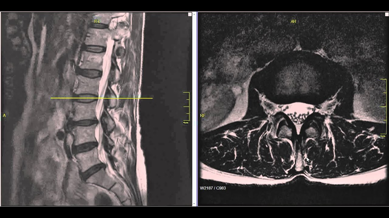
Lumbar Disc Herniation Mri Explained Dr Jeffrey P Johnson Hd Youtube Lumbar Disc Disk Herniation Mri
Pin On Hernie
Color Mri Of The Lumbar Spine Low Back Showing A Normal Lumbar Spine And One With Disc Herniations Created By Medical Media Imag Disk Herniation Mri Anatomy

Pin On Disc Prolapse Slipped Bulged Disc


Posting Komentar untuk "Mri Pictures Of Cervical Herniated Disc"