Ruptured Internal Carotid Artery Aneurysm
The EOCME is accredited by the Accreditation Council for. A decrease in the proportion of patients who achieve complete aneurysm occlusion without significant parent artery stenosis has been observed with the use of the device in the communicating segment C7 of the internal carotid artery 474 919 subjects in the PREMIER study at 1 year including those IAs fed by the posterior circulation or.

Ruptured Internal Carotid Artery Aneurysm A Lateral 2d Dsa Image A Download Scientific Diagram
Brief Reports and Innovations is a gold open access journal launched by Annals of Vascular Surgery.

Ruptured internal carotid artery aneurysm. An aneurysm is an abnormal dilatation of the blood vessel wall that involves all three muscular layers of the vessel. A carotid-cavernous fistula CCF is the result of an abnormal vascular connection between the internal carotid artery ICA or external carotid artery ECA and the venous channels of the cavernous sinus. Aside from the bleeding issues there is significant risk of artery spasm leading to stroke. Aneurysms may be a result of a hereditary condition or an acquired disease. The patient is status post remote craniotomy and clipping of ruptured ACOM aneurysm. The resultant bleeding into the space around the brain is called a subarachnoid hemorrhage SAH.
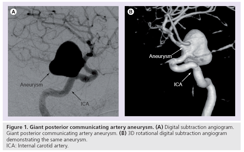
The prevalence of AAA has been reported to range from 2. Although usually heard with the stethoscope such sounds may occasionally also be palpated as a thrill. The internal carotid arteries form the anterior green circulation and the vertebral basilar arteries supply the posterior red circulation of the brain. A cerebral or intracranial aneurysm is an abnormal focal dilation of an artery in the brain that results from a weakening of the inner muscular layer the intima of a blood vessel wall. Posterior communicating artery branch of the internal carotid. Most aneurysms 82 had non-saccular morphology and 56 were giant sized.
The vessel develops a blister-like dilation that can become thin and rupture without warning. Aneurysms can also be a nidus starting point for clot formation and embolizationThe word is from Greek. Most often a ruptured brain aneurysm occurs in the space between the brain and the thin tissues covering the brain. They often occur in areas where the arterial walls are thin or weak. Intraoperative Management of Adult Patients on Extracorporeal Membrane Oxygenation. If untreated another 50 will die within a month with a 20 risk of rebleed by the end of the first two weeks.
The new surgical journal seeks high-quality case reports small case series novel techniques and innovations in all aspects of vascular disease including arterial and venous pathology trauma arteriovenous. An aneurysm occurs when an artery wall becomes weakened and the pressure of blood flow stretches it thin. A type 1 excludes note is a pure excludes. Multiple connections to other key vessels including ophthalmic internal carotid MHT ILT ascending pharyngeal occipital these can be. However at their most severe aneurysms can lead to life-threatening internal bleeding. An aneurysm is a dilation of an artery.
Definition and Epidemiology The word aneurysm is derived from the Greek work aneurusma or widening An aortic aneurysm is an enlargement dilatation of the aorta to greater than 15 times normal size. The basilar artery and the internal carotid arteries communicate with each other in a ring at the base of the brain called the Circle of Willis. Many have no symptoms and are not dangerous. A brain aneurysm can leak or rupture causing bleeding into the brain hemorrhagic stroke. About 10 of patients with a ruptured aneurysm die before receiving medical care.
The Editors of Clinical Imaging in conjunction with the Elsevier Office of Continuing Medical Education are pleased to offer an AMA PRA Category 1 CME credit program for registered Clinical Imaging physician reviewers who complete manuscript reviews. An aneurysm is a weakening and bulging of an artery wall. Annals of Vascular Surgery. The stretching results in a fragile bulge in the artery that if ruptured can lead to fatal internal bleeding. Abdominal Aorto-iliac Artery Aneurysms 2019 Peripheral Arterial Diseases 2018 Atherosclerotic Carotid and Vertebral Artery Disease 2018 Vascular Access 2018 Diseases of Mesenteric Arteries and Veins 2017 Descending Thoracic Aorta Diseases 2017 Chronic Venous Disease 2015. Abdominal aortic aneurysms are usually small and dont cause many noticeable.
Usual origin from the proximal Internal Maxillary Artery IMAX with multiple clinically-important variants. Carotid-cavernous fistula CCF is an abnormal connection between the carotid artery andor its branches and a large vein called the cavernous sinus. This International journal Journal of Clinical Neuroscience publishes articles on clinical neurosurgery and neurology and the related neurosciences such as neuro-pathology neuro-radiology neuro-ophthalmology and neuro-physiology. Computed tomography angiography demonstrated a 114 77 8-cm pseudoaneurysm at the base of the right neck which had arisen from the right subclavian artery and extended into the right superior mediastinum producing significant compression of the right common carotid artery with a tracheal mass effect. Once the artery wall has ruptured it is a medical emergency and the patient is likely to die unless treated swiftly. CCFs are classified based on the arterial.
The journal has a broad International perspective and emphasises the advances occurring in Asia the Pacific Rim region Europe. Occasionally there may be abdominal back or leg pain. A cervical internal carotid artery loop is also present red arrow all of these factors serve to complicate ICA catheterization beyond the aneurysm and decrease stability of embolization setup. A bruit is an audible vascular sound associated with turbulent blood flow. A type 1 excludes note indicates that the code excluded should never be used at the same time as I671A type 1 excludes note is for used for when two conditions cannot occur together such as a congenital form versus an acquired form of the same condition. A carotid-cavernous fistula may be either direct high-flow or spontaneous indirectlow flow.
AMA PRA Category 1 CME credit for Clinical Imaging reviewers. An Expert Consensus Statement From the Society of Cardiovascular AnesthesiologistsPart I Technical Aspects of Extracorporeal Membrane Oxygenation. Treatment of an intracerebral aneurysm is surgical. Basilar strokes are uncommon but may result from a ruptured basilar aneurysm or basilar artery occlusion. An aneurysm is an outward bulging likened to a bubble or balloon caused by a localized abnormal weak spot on a blood vessel wall. The cavernous sinus is located behind the eye and receives blood from brain orbit and pituitary gland.
Common locations included the cavernous internal carotid artery 23 middle cerebral artery 20 and posteroinferior cerebellar artery 12. Symptoms of a ruptured. In the head and neck these auscultatory sounds may originate in the heart cardiac valvular murmurs radiating to the neck the cervical arteries carotid artery bruits the cervical veins cervical venous hum. It often looks like a berry hanging on a stem. Right internal carotid artery aneurysm extracranial portion ICD-10-CM I720 is grouped within Diagnostic Related Groups MS-DRG. Aneurysms most often occur along the aorta.
They usually cause no symptoms except when ruptured. It means not coded here. A brain aneurysm AN-yoo-riz-um is a bulge or ballooning in a blood vessel in the brain.

Left Distal Internal Carotid Artery Ica Aneurysm 18 Mm With Download Scientific Diagram

Asymptomatic Internal Carotid Aneurysm An Uncommon Disease Of The Carotid Arteries Annals Of Vascular Surgery

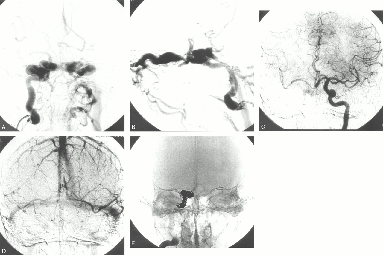
Subarachnoid Hemorrhage Ica Giant

Digital Subtraction Angiogram Of The Left Internal Carotid Artery Download Scientific Diagram

One Aneurysm In The Right Cavernous Segment Of Internal Carotid Artery Download Scientific Diagram

A 53 Year Old Man With Large Left Paraophthalmic Internal Carotid Download Scientific Diagram

Patient 8 A 32 Year Old Man With A 2 Mm Left Middle Cerebral Download Scientific Diagram

Medivisuals Ruptured Left Internal Carotid Artery Aneurysm Medical Illustration

A 38 Year Old Man With Large Ruptured Left Paraophthalmic Internal Download Scientific Diagram
Ruptured Cavernous Sinus Aneurysms Causing Carotid Cavernous Fistula Incidence Clinical Presentation Treatment And Outcome American Journal Of Neuroradiology

Case 2 A 66 Year Old Male Suffered From Sah Caused By Ruptured Right Download Scientific Diagram
Endovascular Treatment Of Intracranial Aneurysms
Aneurysm On The Right Internal Carotid Artery Ica In A 4 Year Old Download Scientific Diagram

Patient No 1 Presented With Headaches A Pre Procedure Right Internal Download Scientific Diagram
Posting Komentar untuk "Ruptured Internal Carotid Artery Aneurysm"