What Are Intracranial Vascular Flow Voids
The glomerulus is composed of a knot of capillaries. Guidelines for the clinical management of patients with neurocysticercosis NCC were prepared by a panel of the Infectious Diseases Society of America IDSA and the American Society of Tropical Medicine and Hygiene ASTMH.
Pin On Snc
It means that there is more fluid in the ventricles and around the cortical sulci.

What are intracranial vascular flow voids. Non-vascular etiologies of pulsatile tinnitus include neoplasm like paraganglioma osseous pathology idiopathic intracranial hypertension and systemic disorders such as anemia 68. If it is associated with neurological symptoms it should be evaluated. Complications like the previous hemorrhage adjacent brain edema and atrophy may be seen. There is subtle adjacent vascular flow voids along the dorsal aspect of the cord. TM is characterized by weakness and. A flow void may be seen because of the hypervascularity.
A fusiform mass with intramural cystic change and a vector of spread that projects upward and medial toward the lateral medulla suggests schwannoma. Stenosis with thickened carotid artery walls is seen in patients with Takayasu disease. The AV shunt is located inside the dura mater close to the spinal nerve root where the arterial blood. Intracranial or carotid artery aneurysm has been reported in patients with polyarteritis nodosa and HIV-related vasculitis 13. Its quite an experience hearing the sound of your voice carrying out to a over 100 first year. A major complication of deep vein thrombosis is pulmonary embolism.
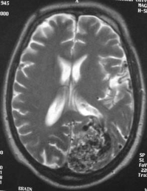
B Fat-saturated contrast-enhanced T1-weighted image shows enhancement. All medical devices in India follow a regulatory framework that is based on the drug regulations under the Drugs and Cosmetic Act 1940 and Drugs and Cosmetic Rules 1945. A b Axial proton-densityweighted a and gadolinium-enhanced T1-weighted b MR images show multiple flow voids and contrast-enhanced tubular structures representing a large vascular lesion that involves the entire right cerebral hemisphere. Spinal dural arteriovenous AV fistulas are the most commonly encountered vascular malformation of the spinal cord and a treatable cause for progressive para- or tetraplegia. They most commonly affect elderly men and are classically found in the thoracolumbar region. However this measure does not assure patency of the device.
B The presence of a bruit indicates good blood flow through the device. Flow voids are best seen on T2-weighted and FLAIR images but can sometimes also be seen on T1-weighted images. Signs and symptoms which may occur suddenly and require immediate treatment include dyspnea severe chest pain apprehension cough possibly accompanied by hemoptysis tachycardia fever hypotension diaphoresis pallor shortness of breath and friction rub. D The nurse should inspect the vascular access site frequently for signs of infection. There are atherosclerotic calcifications fo the intracranial circulation. A thrombus will manifest as absence of flow void.
The Bowman or glomerular capsule surrounds the glomerulus. Transverse implies that the inflammation extends horizontally across the spinal cord. Fast flow in a conglomerate of tangled blood vessels generates serpiginous and tubular flow voids seen on bothT1 and T2 however mostly evident on T2 weighted images. The classification of medical devices rules along with regulatory approval and registration by the CDSCO is under the control of Drug Controller General of India DCGI. C The nurse should inspect the site for bruising or hematoma. On spin-echo images patent cerebral veins usually will demonstrate low signal intensity due to flow void.
Finally the piezoelectric effect is the property of some crystalline materials to polarize when subjected to a mechanical deformation direct piezoelectric effect generating a potential difference and at the same time to deform in an elastic manner when traversed by electrical current inverse piezoelectric effect. Partial transverse myelitis and partial myelitis are terms sometimes used to specify inflammation that only affects part of the width of the spinal cord. Now perhaps the. This usually a normal finding in the elderly and in babies. 48 Likes 2 Comments - College of Medicine Science mayocliniccollege on Instagram. This might be explained by.
The glomerulus derives its blood flow from the renal artery of the abdominal aorta. There is generalized parenchymal volume loss without hydrocephalus midline shift or apparent mass effect. The guidelines are intended for infectious disease specialists neurologists neurological surgeons internists pediatricians and family. Vascular wall thickening with enhancement of the temporal arteries is seen in patients with giant cell arteritis. Cerebral small vessel disease SVD is a frequent finding on CT and MRI scans of elderly people and is related to vascular risk factors and cognitive and motor impairment ultimately leading to dementia or parkinsonism in some. But compatible with chronic microvascular ischemic change.
Transverse myelitis TM is a rare neurological condition in which the spinal cord is inflamed. Answer 1 of 7. The vascular system of the nephron consists of the glomerulus and the Bowman capsule both located in the cortex of the kidney. Pulsatile tinnitus is perceived unilaterally in most cases though it can occur bilaterally in case of systemic vascular disease or the presence of a midline. If the tumor has low-signal high-velocity flow voids with vector of spread through the floor of the middle ear cavity glomus jugulare is the first diagnostic consideration. Catheters placed in the kidney pelvis are irrigated using gentle pressure and small amounts of sterile saline solution or 5 mL at one time to avoid damaging renal tissues.
MR imaging is superior to CT for detecting the intracranial extension which is seen in 1030 of cases. In order to maintain gravity flow the drainage bag should be hung below the level of the bladder. MR imaging shows iso- to hyperintensity on T2-weighted image and hypointensity on T1-weighted image with avid contrast enhancement. There is intrinsic thoracic cord T2 hyperintensity representing edema. This measure does not assess patency. Absence of normal flow void on MR.
Academiaedu is a platform for academics to share research papers. Program within mayoclinicgradschool is currently accepting applications. The flow of urine is dependent on gravity. The normal brain parenchyma is interspersed between the abnormal vessels. In general the relations are weak and not all subjects with SVD become demented or get parkinsonism. Flow-voids are seen in the basilar and internal carotid.
Why Are There Flow Voids In Major Intracranial Vessels

Fd Hydatid Cyst Diagnostic Imaging Bone Diseases Cysts

Cerebral Vascular Malformations Radiology Reference Article Radiopaedia Org Radiology Vascular Celestial Bodies

Trident And Face Of Giant Panda Sign On Mri In Wilsons Disease Note Deposition Of Copper In Basal Ganglia Mri Brain Brain Anatomy Wilson S Disease

Abnormal Flow Voids Without Cord Signal Are Seen In Both Patients With Download Scientific Diagram

Cerebral Vascular Malformations Radiology Reference Article Radiopaedia Org Radiology Sinusitis Vascular

Pin On Neuromedicine

Pin By Dafinka Momcheva On Radiology In 2021 Mri Brain Radiology Ct Scan

Lyre Sign Carotid Artery The Lyre Sign Refers To The Splaying Of The Internal And External Carotid By A Carotid Body Tumour Classi Carotid Artery Tumor Body

Sinus Pericranii Radiology Case Radiopaedia Org Radiology Radiology Imaging Sinusitis

Connatal Cyst Mri Nuclear Medicine Cns Mri

Dermoid Cyst Brain Google Search Radiology Brain Lesions Dermoid Cyst

Brain Imaging In Arteriovenous Malformation
A Mri Showing Patent Functioning And A Flow Void At The Floor Of The Download Scientific Diagram


Posting Komentar untuk "What Are Intracranial Vascular Flow Voids"