Mri Scan For Thigh Pain
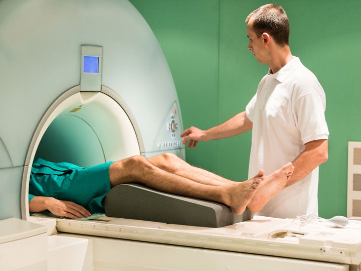
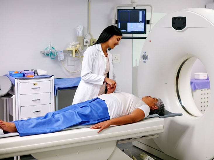
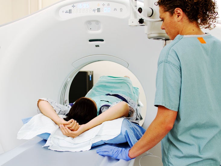
Like a CT scan an MRI scan may be done in a hospital or at an outpatient imaging center. If a stress fracture is suspected but not confirmed a doctor may recommend beginning non-surgical treatment right away.
Hip Arthritis In Crohn S Disease Mri Scan Stock Image C030 2168 Science Photo Library

The conditions previously mentioned are commonly behind the warm sensation in.
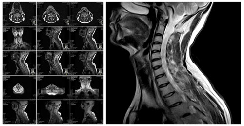
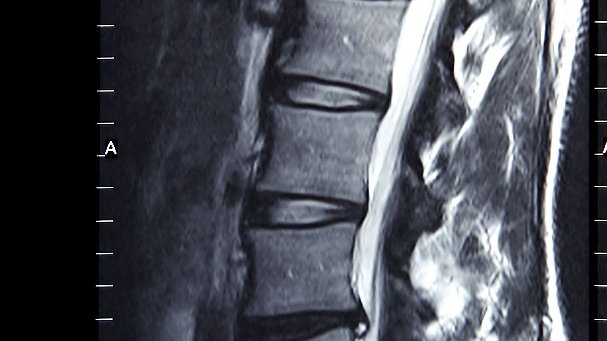
Mri scan for thigh pain. I have had nerve conduction studies tried massage therapy active release therapy acupuncture physiotherapy a Botox injection into the piriformis muscle and a cortisone injection into the ischial tuberosity area where the pain seems to radiate from MRI and ultra sound imaging show no notable abnormalities. MRI cervical spine with IV contrast. A weight is then applied downwards onto the thigh. If your back pain persists for six weeks and does not respond to pain relievers or physical therapy then an MRI scan of the lumbar spine may be appropriate MRIs are frequently overused so dont rush to imaging if your back pain is an isolated ailment. A grade three thigh strain is a complete rupture of a muscle and is a serious. Idiopathic Chondrolysis of the Hip.
What causes dull achy thigh pain. Symtpoms may also be referred into the knee. Most folk have heard of sciatica and the nerve which comes from the lower lumbar spine. May Be Appropriate Disagreement CT cervical spine without and with IV contrast Usually Not Appropriate FDG -PETCT skull base to mid -thigh. Plain films are usually negative early in the course of fracture. It supplies the back of the thigh side of the lower leg and much of the foot.
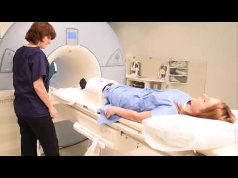
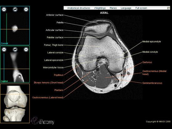
Pain is likely to be felt when stretching the hamstring muscles and becomes worse with exercise particularly repetitive exercises like long-distance running. This webpage presents the anatomical structures found on thigh MRI. If pain is reproduced then the test is positive and it may be a femur stress fracture. It is confirmed by pain on stretch and contraction of the muscle. It may take from 30 to 60 minutes to do the study. You may get x-rays a CT scan or an MRI.
While pain and discomfort are sometimes unavoidable arming yourself with accurate information can help you feel your best and prevent unnecessary complications. May Be Appropriate Disagreement O Radiography cervical spine. Pressing down on the skin over the hip may cause pain. A grade two Thigh strain is usually sore to touch. With a grade two thigh strain there is immediate pain which is more severe than the pain of a grade one injury and produces pain on walking. While you lie face up on the table the doctor puts one hand under your buttock while bending your hip and knee at a 90-degree angle.
The femur is the longest and strongest bone in the body. Dear doctor since 3 years I have been suffering from backacheOn 24th February 2011 I took an MRI scan of lumbar spine. Thigh pain symptoms can develop acutely or can be a chronic problem that worsens over time and they can occur in different parts of the thigh like the front back or side. Pain commonly experienced in the anterior thigh. Brain magnetic resonance imaging MRI is a common medical imaging method that allows clinicians to examine the brains anatomy1. The thigh has some of the bodys largest muscles.
What is a stress fracture of the femur. This measures the electrical activity in your muscles through a thin needle electrode. Traumatic shoulder pain is shoulder pain believed to be directly attributed to a traumatic event either acute or chronic. Stress fractures occur when muscles are too exhausted to absorb shock. The Human Gene Mutation Database. MRI BIOEFFECTS SAFETY AND PATIENT MANAGEMENT is a comprehensive authoritative textbook on the health and safety concerns of MRI technology that contains contributions from more than forty internationally respected experts in the field.
Modic End Plate Changes of Spine With Classification. An X-ray may or may not show up the stress fracture but a bone scan or MRI should give a more accurate diagnosis. Bone scan or MRI are the test of choice. Poorly defined condition in which articular cartilage of the hip is injured by an undefined inflammatory process. Patients reported pain on the side and front part of their thighs A buzzing sensation inside the thigh and muscle numbness is also very commonly felt. At the Institute of Medical Genetics in Cardiff.
Image_slider Thigh Magnetic Resonance Imaging. It uses a magnetic field and radio waves to produce detailed images of the brain and the brainstem to detect various conditions2. At L1-L2 L2-L3 and L3-L4 no disc desiccation seen. Its result is shown below. See All About Stress Fractures. MRI can detect tumors infections and disk herniation and it can assess for edema in the pars interarticularis region which is consistent with acute spondylolysis12 14 A.
This pain may be the result of either fracture the clavicle scapula or proximal humerus or soft-tissue injury most commonly of the rotator cuff acromioclavicular ligaments or labroligamentous complex. An MRI may be used to help diagnose torn knee ligaments and cartilage torn rotator cuffs herniated disks osteonecrosis bone tumors and other problems. If you have meralgia paresthetica the results. Usually Not Appropriate Bone scan whole body with SPECT or SPECTCT neck. This suspicion can be confirmed through a bone scan or MRI. Thigh pain can have a variety of causes ranging from acute injury to.
FM usually in second decade of life. However the pain will subside when sitting. The bone of the thigh is called the femur. Femoral nerve damage causes severe pain in the buttock and upper anterior thigh and lower inner leg pain. The patient may experience a gradual onset of deep buttock or thigh pain following a sprinting session. The usual sites for this kind of fracture are the femur and the pubic bone.
Thigh pain is a common injury but that does not mean it cant be serious. Damage disruption or injury to any of its components can result in dully achy thigh pain. A definitive diagnosis may require advanced imaging such. Thigh muscles are responsible for allowing normal gait and proper lower extremity function1. If further assessment is required an MRI scan X-ray or ultrasound may be used to confirm the severity. The thigh contains one major bone and many muscles nerves and arteries.
This page was updated by Dr Barrie Lewis on 29th October 2018. If the patient reports worsening pain when he or she puts weight on one leg there is even more of a chance that there is a stress fracture. Ganesh Diagnostic is a renowned diagnostic centres and pathology labs Rohini in the state of Delhi in India. It offers diagnostic and imaging services such as ultrasound x. It serves as the definitive resource for radiologists and other physicians MRI technologists physicists.
Knee Mri Scan Protocols Positioning And Planning Youtube
How Long Does A Knee Mri Take Aica Orthopedics

Knee Mri Scan Purpose Procedure And Risks
Can You See Nerve Damage In An Mri
Lumbar Mri Scan Purpose Procedure And Risks
Pin On Excalibur Healthcare S Imaging Teleradiology Pins

An Mri Scan Of His Pelvis Left And Right Thigh With And Without Download Scientific Diagram

Pelvis Mri Scan Risks Preparation And Procedure
Disc Sequestration Radiology Imaging Radiology Mri

Mri Knee Google Search Mri School Radiology Mri
How Much Does It Cost For An Mri Of The Leg Affordablescan

Knee Joint Mri Scan Stock Image C002 1301 Science Photo Library

Indications And Contraindications For An Mri Scan
Diagnostic Tests X Ray Ct Scan Mri


Posting Komentar untuk "Mri Scan For Thigh Pain"